Review
. 2018 Jan 1:123:33-64.
doi: 10.1016/j.addr.2017.08.001. Epub 2017 Aug 4.

Nanomedicine and advanced technologies for burns: Preventing infection and facilitating wound healing

Affiliations
Review

Nanomedicine and advanced technologies for burns: Preventing infection and facilitating wound healing

Mirza Ali Mofazzal Jahromi et al. Adv Drug Deliv Rev. .

Abstract

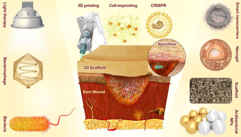
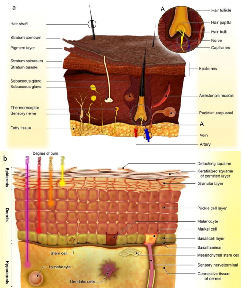
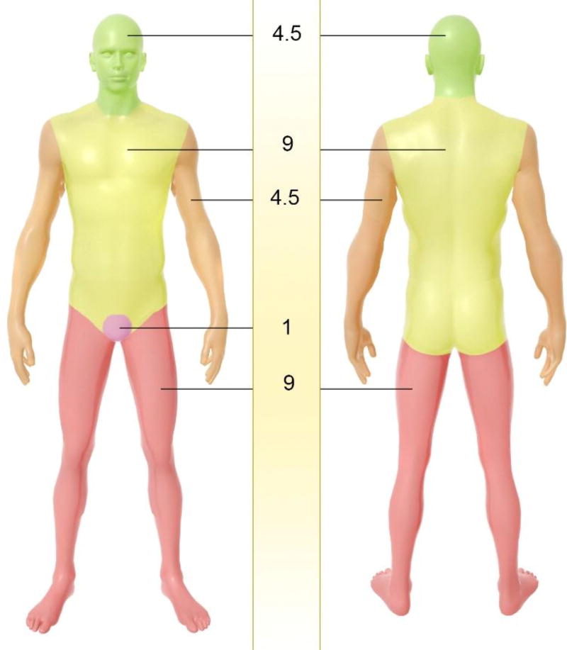
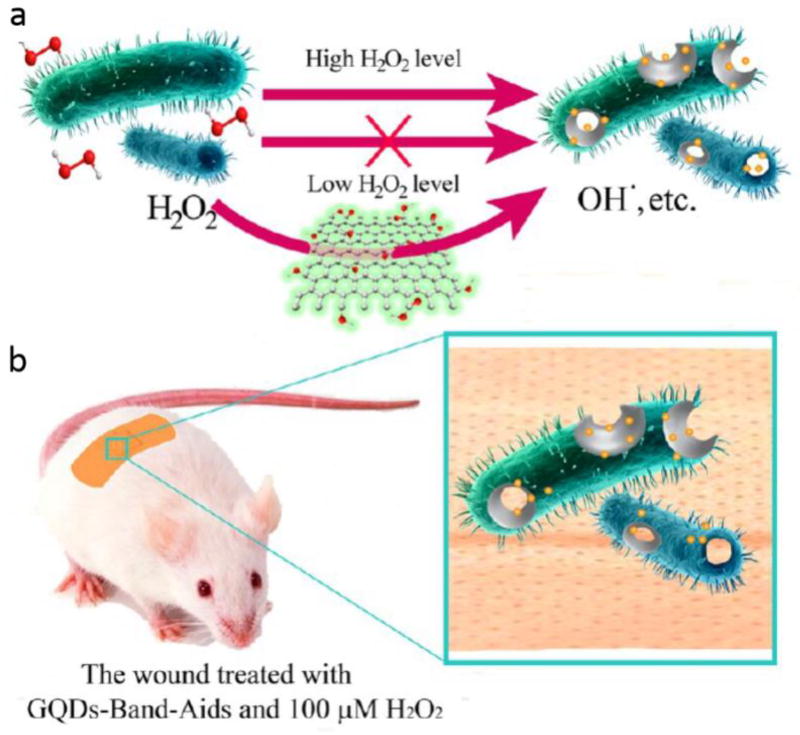
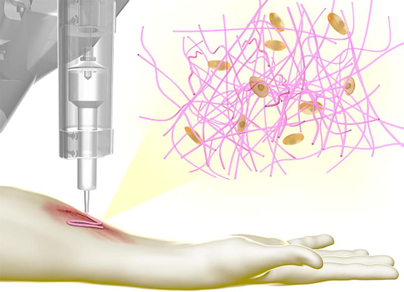
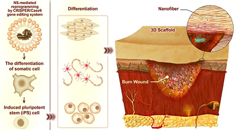
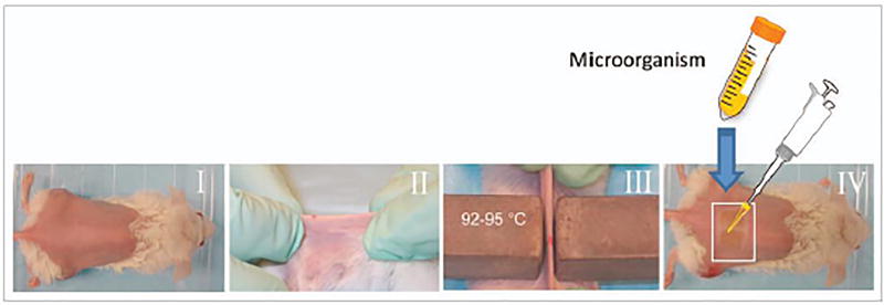
According to the latest report from the World Health Organization, an estimated 265,000 deaths still occur every year as a direct result of burn injuries. A widespread range of these deaths induced by burn wound happens in low- and middle-income countries, where survivors face a lifetime of morbidity. Most of the deaths occur due to infections when a high percentage of the external regions of the body area is affected. Microbial nutrient availability, skin barrier disruption, and vascular supply destruction in burn injuries as well as systemic immunosuppression are important parameters that cause burns to be susceptible to infections. Topical antimicrobials and dressings are generally employed to inhibit burn infections followed by a burn wound therapy, because systemic antibiotics have problems in reaching the infected site, coupled with increasing microbial drug resistance. Nanotechnology has provided a range of molecular designed nanostructures (NS) that can be used in both therapeutic and diagnostic applications in burns. These NSs can be divided into organic and non-organic (such as polymeric nanoparticles (NPs) and silver NPs, respectively), and many have been designed to display multifunctional activity. The present review covers the physiology of skin, burn classification, burn wound pathogenesis, animal models of burn wound infection, and various topical therapeutic approaches designed to combat infection and stimulate healing. These include biological based approaches (e.g. immune-based antimicrobial molecules, therapeutic microorganisms, antimicrobial agents, etc.), antimicrobial photo- and ultrasound-therapy, as well as nanotechnology-based wound healing approaches as a revolutionizing area. Thus, we focus on organic and non-organic NSs designed to deliver growth factors to burned skin, and scaffolds, dressings, etc. for exogenous stem cells to aid skin regeneration. Eventually, recent breakthroughs and technologies with substantial potentials in tissue regeneration and skin wound therapy (that are as the basis of burn wound therapies) are briefly taken into consideration including 3D-printing, cell-imprinted substrates, nano-architectured surfaces, and novel gene-editing tools such as CRISPR-Cas.

Keywords: 3D printing; Burn wound infection; CRISPR; Cell-Imprinting; Gene therapy; Growth factors; Nanomedicine; Nanoparticles; Stem cells; Stimulus-responsive drug delivery; Topical treatment; Wound healing.


Figures

Figure. 1
Figure. 1
Schematic illustration of important conventional, developing or innovative approaches, (such as light therapy; therapeutic microorganisms e.g. bacteriophages and bacteria with bactericidal effects; antibacterial NPs or NPs with therapeutic effects e.g. silver NPs and gold NPs; innovative nanocarriers e.g. smart nanocarriers and nanogels; novel scaffolds for wound dressing, (stem) cell therapy and regenerative medicine, therapeutic (drug, growth factor, gene, etc.) delivery, and so forth; and innovative technologies e.g. 3D-printing, cell-imprinting, and CRISPR-Cas9 gene editing), which have been the subject of various researches, conducted with the aim of wound healing and prevention of infection in burns.
Figure 2
Figure 2
Schematic illustration of: a) the structure of skin, b) layers of skin including epidermis, dermis and hypodermis, and their constituent cells and sub-layers.
Figure 3
Figure 3
Assessment of skin burn in terms of total body surface area (TBSA) percent, using the rule of nines in applied area, thus to quickly estimate the affected TBSA percent.
Figure 4
Figure 4
a) The platform based on GQDs/low level H2O2 used as antibacterial agent, b) The GQD equipped bandage for disinfection of the wound site in vivo Reprinted with permission from ref. [239], copyright 2014, American Chemical Society.
Figure 5
Figure 5
Curves showing morphological measurements evaluations from mice treated with SHAM group, GEL (hydrogel), GEL/ASCs at the burn wound site: a) wound size measurement; 1, 2, and 3 weeks after burn injury initiation compared to initial injury. Here, no effect of the gel wound dressing and wound closure was reported, b) and c) wound thickness (mm) after 1 and 2 week, and area occupied by nuclei, respectively, indicative of granulation tissue, which was facilitated by ASC-containing gels, c) Quantitative measurement of blood vessels in regenerating regions 1 week after the burn initiation. SHAM group (untreated), GEL group (treated only with P-fibrin gel), and GEL/ASCs group (GEL group integrated with adipose-derived stem cells) Reprinted with permission from ref. [267], copyright 2016, John Wiley & Sons, Inc.
Figure 6
Figure 6
Schematic of the synthesis procedure of QCSP/PEGS-FA hydrogels. Reprinted with permission from ref. [277], copyright 2017, Elsevier.
Figure 7
Figure 7
a) Schematic illustrating the fabrication process of the biocomposite wound dressing, b) microscopic images of 8mm full thickness wounds of C57BL/6J mice on day 21 after the burn injury initiation: (i) Spontaneous healing, (ii) OP-Gel, (iii) OP-Gel-NS, (iv) OP-Gel-Cipro, and (v) Bactigras, c) Histological analysis of the excised tissue at wound site: (i) intact tissue at first day, treated tissue after 3 weeks under different conditions of: (ii) Spontaneous healing, (iii) using OP-Gel, (iv) using OP-Gel- NS, (v) using OP-Gel-Cipro and (vi) using Bactigras (commercially available dressing). Reprinted with permission from ref. [278], copyright 2016, Elsevier.
Figure 8
Figure 8
a) Scanning electron microscopy of RADA16, (b) RADA16-FPG and (c) RADA16-RGD. (d) Cell viability result for the implemented keratinocyte (i.e. HaCaT) and fibroblast (i.e. NIH/3T3) using the three 1% w/v RADA16 NF scaffolds and cell control after 0 hour and 3 days of incubation. Reprinted from ref. [288], copyright 2014, Nature (licensed under a Creative Commons Attribution-NonCommercial-NoDerivs 4.0 International License).
Figure 9
Figure 9
a) Fluorescence micrographs of cells showing clustered growth after days 3 on HEMA (left; scaffolds coated with fibronectin were seeded with NIH/3T3 mouse fibroblasts); with homogenous growth of the cells cultured on pH-sensitive DMAEMA/HEMA scaffolds (right) [299], b) percentage swelling of zinc ion containing hydrogels; c) TA release from the hydrogels, at various pH values Reprinted with permission from ref. [300], copyright 2016, American Chemical Society; d) Scheme illustrating the synthesis of enzyme-sensitive self-assembled polymeric vehicles (from PEG-b-PP and PEG-b-PC) for selective delivery of antibiotics. They were designed to be responsive to penicillin G amidase (side chain cleavage) and to β-lactamase (microstructural transformation). This stimulus-responsive nanoplatform resulted in the constant cargo liberation and bioactivity of the encapsulated antimicrobial agent Reprinted with permission from ref. [302], copyright 2015, John Wiley & Sons, Inc.
Figure 10
Figure 10
a) Images of the infected wounds with different treatments: (i, ii) Untreated wounds on control at postoperative day 0 and 7. (iii, iv) Infected untreated wounds on postoperative day 0 and 7. (v, vi) Infected wounds treated ultrasound-activated TiO2 (UIT) on postoperative day 0 and 7, b) Effect of ultrasound-activated TiO2 on the treatment of the infected wound in regard to wound size percentage according to the initial wound size (day 0) Reprinted with permission from ref. [304] copyright 2016, John Wiley & Sons, Inc., c) Scheme of the smart antimicrobial system. Here, the exposure to lysozyme led to hydrolysis of NAc-CTS to give COS. The generated COS served as a substrate for CDH that produces the antimicrobial H2O2. Reprinted with permission from ref. [305], copyright 2017, John Wiley & Sons, Inc.
Figure 11
Figure 11
a) Scheme of a PEG-MoS2 nanoplatform to destroy bacteria via a synergistic combination of peroxidase catalysis and photothermal activation: (i) Capture of the nanoplatform by bacteria; (ii) catalytic activity of the nanoplatform decomposes low-concentration H2O2, leading to generation of ·OH that damages cell wall integrity; 808 nm laser treatment induces hyperthermia, causing accelerated GSH oxidation, b) Relative bacterial viability for B. subtilis, obtained after incubation with PBS, MoS2 (100 µg mL−1), H2O2 (100 µM), or MoS2+H2O2 for 20 min, with or without the laser treatment, c) GSH loss after heating by water bath or 808 nm NIR-irradiation compared to control (non-treated GSH) for 20 min at 50 °C. Reprinted with permission from ref. [315], copyright 2016, American Chemical Society, d) Photomicrographs indicating PDT-triggered healing: (i) normal skin, (ii) wound without infection, (iii) infected wounds, (iv) infected wounds with 200 µM pl–cp6 treatment, and (v) infected wounds with 200 µM pl–cp6 treatment plus with exposure to the light dose of 60 J/cm2. Reprinted with permission from ref. [316], copyright 2013, Springer.
Figure 12
Figure 12
a) Scheme illustrating the preparation of hydrogel sheet loaded with hEGF, used for healing of full thickness wounds, b) Wound closure-time curve in mice; and c) Macroscopic images of wound sites, obtained for different samples compared to non-treated controls. Reprinted with permission from ref. [328], copyright 2016, Elsevier.
Figure 13
Figure 13
Schematic of 3D-printing technology developed for the fabrication of 3D-scaffolds and dressings, within which cell-matrix components can be implanted, with applications in burn wound healing and tissue regeneration.
Figure 14
Figure 14
Schematic representing the concept of using gene editing tools such as CRISPR-Cas for reprogramming of stem cells (e.g. iPSCs) in wound healing applications
Figure 15
Figure 15
I–II) In Stevens burn model, a deep burn wound is generated using two blocks of brass with pre-heating of 92–95°C on a shaved mouse back. III) Square show burned region. IV) Bacterial suspension in PBS was used on the surface of burn. Reprinted from ref. [391], copyright 2011, Taylor and Francis Group (open access, licensed under a Creative Commons Attribution-NonCommercial 3.0 Unported License.)

References

    1. Peck M, Molnar J, Swart D. A global plan for burn prevention and care. Bulletin of the World Health Organization. 2009;87:802–803. - PMC - PubMed
    1. http://www.who.int.

    1. Church D, Elsayed S, Reid O, Winston B, Lindsay R. Burn wound infections. Clinical microbiology reviews. 2006;19:403–434. - PMC - PubMed
    1. Salmon JK, Armstrong CA, Ansel JC. The skin as an immune organ. West J Med. 1994;160:146–152. - PMC - PubMed
    1. Rowan MP, Cancio LC, Elster EA, Burmeister DM, Rose LF, Natesan S, Chan RK, Christy RJ, Chung KK. Burn wound healing and treatment: review and advancements. Critical care. 2015;19:243. - PMC - PubMed

Publication types

Substances