Review
. 2017 Oct 3;56(39):5202-5209.
doi: 10.1021/acs.biochem.7b00443. Epub 2017 Aug 7.

Going Deeper: Biomolecular Tools for Acoustic and Magnetic Imaging and Control of Cellular Function

Affiliations
Review

Going Deeper: Biomolecular Tools for Acoustic and Magnetic Imaging and Control of Cellular Function

Dan I Piraner et al. Biochemistry. .

Abstract

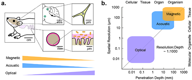
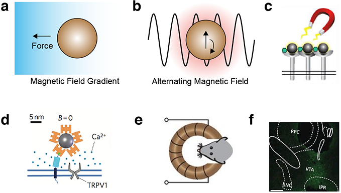
Most cellular phenomena of interest to mammalian biology occur within the context of living tissues and organisms. However, today's most advanced tools for observing and manipulating cellular function, based on fluorescent or light-controlled proteins, work best in cultured cells, transparent model species, or small, surgically accessed anatomical regions. Their reach into deep tissues and larger animals is limited by photon scattering. To overcome this limitation, we must design biochemical tools that interface with more penetrant forms of energy. For example, sound waves and magnetic fields easily permeate most biological tissues, allowing the formation of images and delivery of energy for actuation. These capabilities are widely used in clinical techniques such as diagnostic ultrasound, magnetic resonance imaging, focused ultrasound ablation, and magnetic particle hyperthermia. Each of these modalities offers spatial and temporal precision that could be used to study a multitude of cellular processes in vivo. However, connecting these techniques to cellular functions such as gene expression, proliferation, migration, and signaling requires the development of new biochemical tools that can interact with sound waves and magnetic fields as optogenetic tools interact with photons. Here, we discuss the exciting challenges this poses for biomolecular engineering and provide examples of recent advances pointing the way to greater depth in in vivo cell biology.


Conflict of interest statement

COMPETING INTERESTS

The authors declare no competing financial interests.

Figures

Figure 1 -
Figure 1 -. Modalities for in vivo imaging and control of cellular function.
(a) Diagram of the length scales of several biological processes of interest in vivo, and the degree to which these length scales are accessible by imaging technologies. (b) Approximate length scales and maximal tissue penetration depths accessible by optical, acoustic, or magnetic imaging.
Figure 2 -
Figure 2 -. Biomolecular tools for ultrasound imaging.
(a) Illustration of sound propagation in the imaging medium and received echo used to form the ultrasound image. (b) GVs are hollow protein nanostructures that freely allow diffusion of dissolved gas through their shell but exclude water. GVs are encoded by operons consisting of 814 genes. (c) Representative transmission electron micrograph of purified GV from Halobacterium. (d) Simulation illustrating nanoscale deformation of GVs under ultrasound leading to nonlinear backscattered echo. (e) Amplitude-modulation pulse sequence reveal GVs in mouse colon (Reprinted from Maresca, D et al (2017). Nonlinear ultrasound imaging of nanoscale acoustic biomolecules. Applied Physics Letters, 110(7), 73704, with the permission of AIP Publishing). (f) Multiplexed imaging of genetically engineered GVs. (g) Heterologous expression of GVs in E. coli using an optimized GV gene cluster. (h) Ultrasound image of E. coli expressing GVs or the non-echogenic luminescence reporter, luciferase. (i) Photoacoustic imaging of tumor expressing tyrosinase, and surrounding blood vessels.
Figure 3 -
Figure 3 -. Biomolecular tools for acoustic control.
(a) Ultrasound can be focused at depth in tissue and apply several forms of energy to interface with cells. (b) Schematic of the gene circuit utilized to gate a GFP reporter gene with a temperature-sensitive repressor (TSR), and a panel of tuned variants of temperature-sensitive repressors. (c) MRI thermometry imaging demonstrates a spatial temperature gradient induced by FUS on a plate of bacterial cells, resulting in spatially targeted gene expression. (d) E. coli were injected into both hindlimbs of a nude mouse; after FUS application to the right hindlimb, reporter gene expression is significantly enriched at the site of heating. (e) Diagram of mechanism by which microbubble cavitation can result in membrane deformation leading to mechanoreceptor activation. (f) Microbubbles attached to cultured retinal pigment epithelium cells. (g) Uptake of membrane-impermeable dye into retinal pigment epithelium cells expressing MscL and functionalized with microbubbles. (h) C. elegans worm motor response to ultrasound in a bath of microbubbles.
Figure 4 -
Figure 4 -. Biomolecular tools for magnetic resonance imaging.
(a) Metalloproteins interact magnetically with aqueous 1H nuclear spins, leading to T1 or T2 MRI contrast. (b) Migrating neuroblasts expressing ferritin produce a hypointense track (arrow) in T2 weighted MRI. Asterisks denote adenovirus injection sites. (c) Overexpression of aquaporin enhances passive diffusion of water across the cell membrane, resulting in contrast on diffusion weighted MRI. (d) AQPi expression in mouse xenograft shows significant contrast compared to contralateral GFP expressing xenograft after expression is induced with doxycycline. (e) GVs interact with hyperpolarized xenon dissolved in biological media, producing contrast in 129Xe MRI. (f) Genetically distinct GVs produce different chemical shifts in 129Xe MRI, enabling multiplexed imaging.
Figure 5 -
Figure 5 -. Biomolecular tools for magnetic control.
(a) Magnetic field gradients exert a force on magnetic particles. (b) Alternating magnetic fields heat magnetic particles by inducing oscillations in the magnetic moment of the nanoparticle. (c) Magnetic fields can induce the clustering of magnetic particles, and receptors to which they are bound. (d) Coupling magnetic nanoparticles to the heat-sensitive ion channel TRPV1 enables magnetic control of calcium influx to the cell. (e) Remote deep brain stimulation using alternating magnetic fields applied to mice with implanted magnetic nanoparticles and virally transduced TRPV1. (f) Expression of the neural activity marker cFos in the ventral tegmental area (VTA) of the mouse brain after injection with TRPV1-encoding virus, magnetic nanoparticles and the application of alternating field stimulation.

References

    1. Donaldson GP, Lee SM, and Mazmanian SK (2015) Gut biogeography of the bacterial microbiota. Nat. Rev. Microbiol 14, 20–32. - PMC - PubMed
    1. Foster FS, Pavlin CJ, Harasiewicz KA, Christopher DA, and Turnbull DH (2000) Advances in ultrasound biomicroscopy. Ultrasound Med. Biol 26, 1–27. - PubMed
    1. Errico C, Pierre J, Pezet S, Desailly Y, Lenkei Z, Couture O, and Tanter M (2015) Ultrafast ultrasound localization microscopy for deep super-resolution vascular imaging. Nature 527, 499–502. - PubMed
    1. Humphrey VF (2007) Ultrasound and matter-Physical interactions. Prog. Biophys. Mol. Biol 93, 195–211. - PubMed
    1. Pankhurst QA, Connolly J, Jones SK, and Dobson J (2003) Applications of magnetic nanoparticles in biomedicine. J. Phys. D. Appl. Phys 36, R167–R181.

Publication types