. 2017 Sep 5;56(35):4722-4731.
doi: 10.1021/acs.biochem.7b00616. Epub 2017 Aug 23.

Regulation of Focal Adhesion Kinase through a Direct Interaction with an Endogenous Inhibitor

Affiliations

Regulation of Focal Adhesion Kinase through a Direct Interaction with an Endogenous Inhibitor

Taylor J Zak et al. Biochemistry. .

Abstract

Focal adhesion kinase (FAK) plays a key role in integrin and growth factor signaling pathways. FAK-related non-kinase (FRNK) is an endogenous inhibitor of FAK that shares its primary structure with the C-terminal third of FAK. FAK S910 phosphorylation is known to regulate FAK protein-protein interactions, but the role of the equivalent site on FRNK (S217) is unknown. Here we determined that S217 is highly phosphorylated by ERK in cultured rat aortic smooth muscle cells. Blocking phosphorylation by mutation (S217A) greatly increased FRNK inhibitory potency, resulting in strong inhibition of FAK autophosphorylation at Y397 and induction of smooth muscle cell apoptosis. FRNK has been proposed to compete for FAK anchoring sites in focal adhesions, but we did not detect displacement of FAK by WT-FRNK or superinhibitory S217A-FRNK. Instead, we found FRNK interacted directly with FAK, and this interaction is markedly strengthened for the superinhibitory S217A-FRNK. The results suggest that FRNK limits growth and survival signaling pathways by binding directly to FAK in an inhibitory complex, and this inhibition is relieved by phosphorylation of FRNK at S217.


Conflict of interest statement

Conflict of interest: The authors declare that they have no conflicts of interest with the contents of this article.

Figures
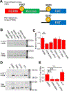
Fig. 1.
Fig. 1.. FRNK serine 217 is highly phosphorylated by ERK.
(A) Schematic cartoon comparing FAK to FRNK including the phosphorylation sites tyrosine 397 and serine 910 (217 on FRNK) (B) Western blot of RASMs treated with various kinase inhibitors probed with P-S217 or total FRNK antibodies. (C) Quantification of (B) for N = 4 biological replicates revealing that ERK inhibition reduced FRNK S217 phosphorylation. (D) Kinase inhibitors reduced FRNK S217 phosphorylation while angiotensin II stimulation resulted in no significant increase in phosphorylation. UI = uninfected cells. (E) Quantification of (D) for N = 3 biological replicates show FRNK is highly phosphorylated at position S217 by ERK. * indicates p < 0.05 and ** indicates p < 0.005.
Fig. 2.
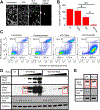
Fig. 2.. S217A-FRNK is a super inhibitory mutant with respect to FAK Y397 phosphorylation and survival.
(A) Fluorescence microscopy of cells infected with a control virus (GFP), WT-FRNK, or S217A-FRNK and stained with DAPI. (B) Quantification of A for N = 4 biological replicates demonstrated that apparent cell survival was strongly reduced by S217A-FRNK compared to WT-FRNK. Data shown are mean ± SE. (C) RASMs expressing β-gal, Cer-FRNK, or CFP-S217A-FRNK, were subjected to flow cytometry apoptosis analysis. Double negative cells indicate viable cells (V). Annexin V positive, PI negative cells indicate apoptotic cells (A), and PI positive, Annexin V negative cells indicate dead cells (D). The data show that the non-phosphorylatable FRNK has increased potency for inducing apoptosis compared to wild type FRNK. (D) Western Blots of RASMs expressing wild type mCherry-FRNK (WT-FRNK) or mutant CFP-S217A-FRNK (S217A-FRNK) were probed with FAK P-Y397 antibodies or total FRNK antibodies. Increasing expression of FRNK resulted in decreased FAK phosphorylation at activation site Y397. Red boxes identify samples with similar expression of FRNK; these lanes are reproduced at right (E) for side-by-side comparison. UI = uninfected cells. GAPDH was used as a loading control. * indicates p < 0.05 and ** indicates p < 0.005.
Fig. 3.
Fig. 3.. FRNK does not displace FAK from focal adhesions, as measured by TIRF microscopy.
Fluorescence microscopy of cells infected with WT-FRNK (A) or S217A-FRNK (B) and co-infected with YFP-FAK. YFP-FAK localized strongly to focal adhesions whether they contained FRNK (cyan arrow) or not (yellow arrow). (C) Quantification of FAK anchoring in focal adhesions (N=74) demonstrated that increasing FRNK expression did not decrease FAK focal adhesion localization, nor was there a significant difference between WT-FRNK and superinhibitory S217A-FRNK.
Fig. 4.
Fig. 4.. FRNK co-immunoprecipitates with FAK.
RASMs infected with Cer-WT-FRNK or CFP-S217AFRNK were lysed in a non-denaturing buffer and endogenous FAK was immunoprecipitated with a FAK-specific antibody that does not recognize FRNK. FAK and co-immunoprecipitated FRNK were detected on the blot using a different antibody that is pan-specific, recognizing both FAK and FRNK. S217A-FRNK exhibited approximately five-fold increased co-immunoprecipitation with FAK compared to wild type. GAPDH was used as a loading control.
Fig. 5.
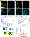
Fig. 5.. FAK and FRNK form oligomeric complexes.
(A, B) TIRF microscopy revealed YFP-FAK localization in focal adhesions in cells coexpressing WT- or S217A-FRNK. Acceptor-selective photobleaching of YFP-FAK resulted in increased fluorescence of non-phosphorylatable FRNK, but not WT-FRNK. (C, D) Quantification of acceptor photobleaching experiments (N=24) for WT- or S217AFRNK coexpressed with YFP-FAK. Red arrows indicate initiation of photobleaching. Data shown are mean ± SE. (E) FRET (black arrows) from a donor (“D”) is abolished after photobleaching of the conjugate acceptor (“A”) in a dimer, resulting in increased donor brightness. For a higher order oligomer, FRET persists until all acceptors have been bleached. (F) The data in (C, D) plotted as donor vs. acceptor intensity revealed significant deviation from a linear relationship (grey line) for non-phosphorylatable FRNK, suggesting multiple acceptors in complex with each donor. The green arrow denotes the direction of change over time. Data are mean ± SE.
Fig. 6.
Fig. 6.. Summary model of FAK activation.
FAK translocates from the cytoplasm to focal adhesions where it forms homo-oligomers. Activated by diverse stimuli, FAK autophosphorylates at Y397, activating growth, survival, and migration signaling pathways. Unchecked activity can lead to pathological conditions such as cancer or excessive neointimal formation as seen in restenosis. FRNK interacts directly with FAK, forming an inhibitory hetero-oligomeric complex in the focal adhesion. Inhibition is relieved by ERK phosphorylation of FRNK on S217.

References

    1. Govindarajan G, Eble DM, Lucchesi PA, and Samarel AM (2000) Focal adhesion kinase is involved in angiotensin II-mediated protein synthesis in cultured vascular smooth muscle cells, Circ. Res 87, 710–716. - PubMed
    1. Heidkamp MC, Bayer AL, Kalina JA, Eble DM, and Samarel AM (2002) GFP-FRNK disrupts focal adhesions and induces anoikis in neonatal rat ventricular myocytes, Circ. Res 90, 1282–1289. - PubMed
    1. Walkiewicz KW, Girault JA, and Arold ST (2015) How to awaken your nanomachines: Site-specific activation of focal adhesion kinases through ligand interactions, Prog. Biophys. Mol. Biol 119, 60–71. - PubMed
    1. Parsons JT (2003) Focal adhesion kinase: the first ten years, J. Cell Sci 116, 1409–1416. - PubMed
    1. Mitra SK, Hanson DA, and Schlaepfer DD (2005) Focal adhesion kinase: in command and control of cell motility, Nat. Rev. Mol. Cell Biol 6, 56–68. - PubMed

Publication types

MeSH terms

Substances

LinkOut - more resources