. 2017 Aug 7;9(8):210.
doi: 10.3390/v9080210.

Viral Ubiquitin Ligase Stimulates Selective Host MicroRNA Expression by Targeting ZEB Transcriptional Repressors

Affiliations

Viral Ubiquitin Ligase Stimulates Selective Host MicroRNA Expression by Targeting ZEB Transcriptional Repressors

Gabriel Lutz et al. Viruses. .

Abstract

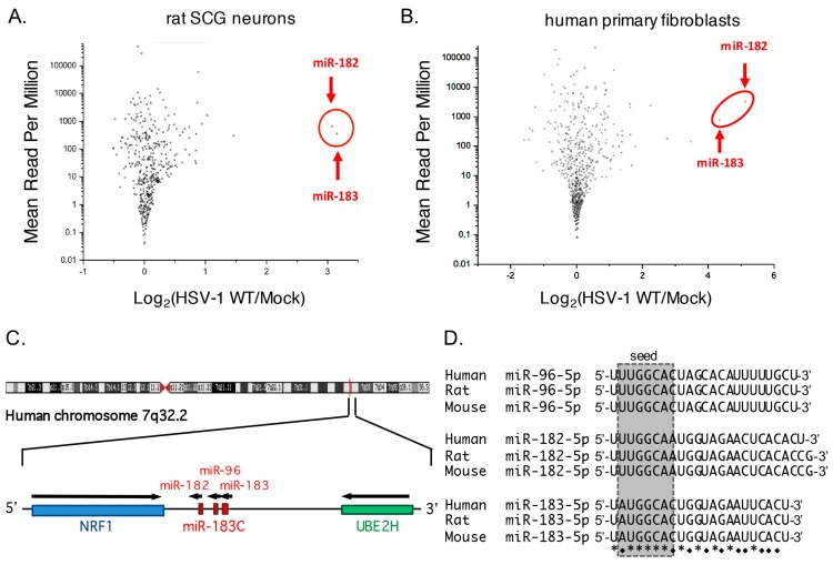
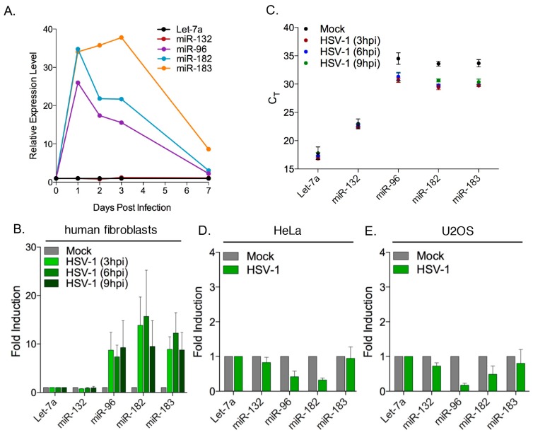
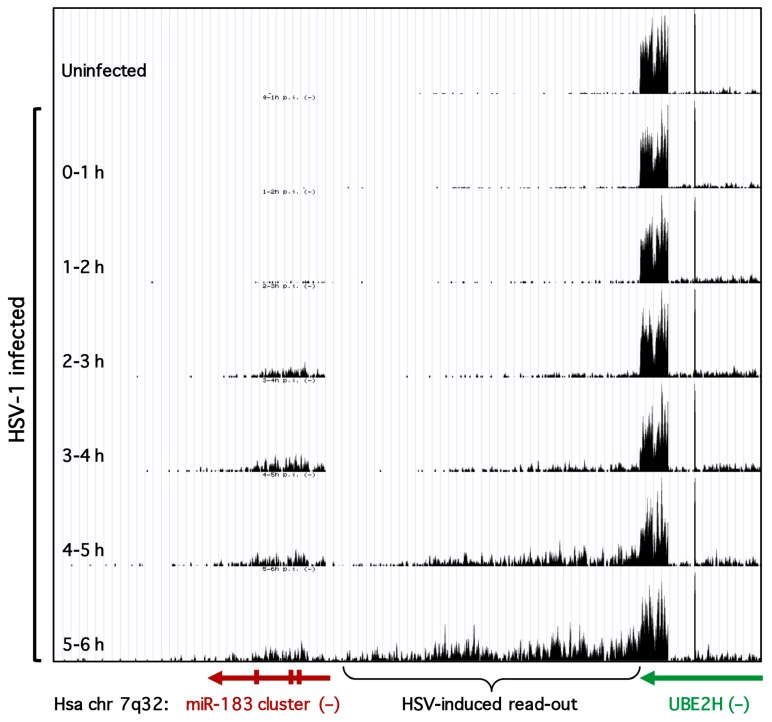
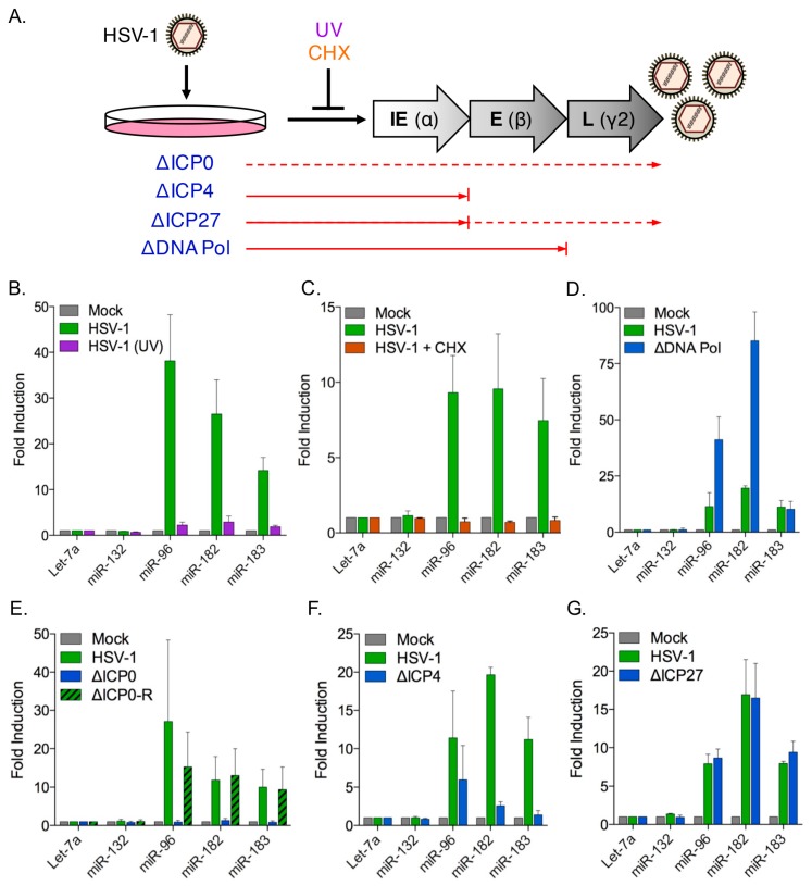
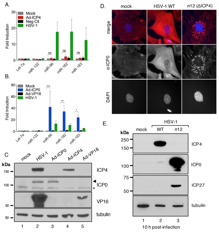
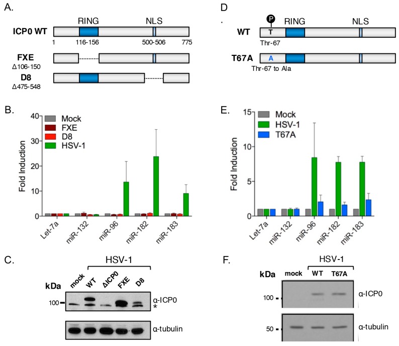
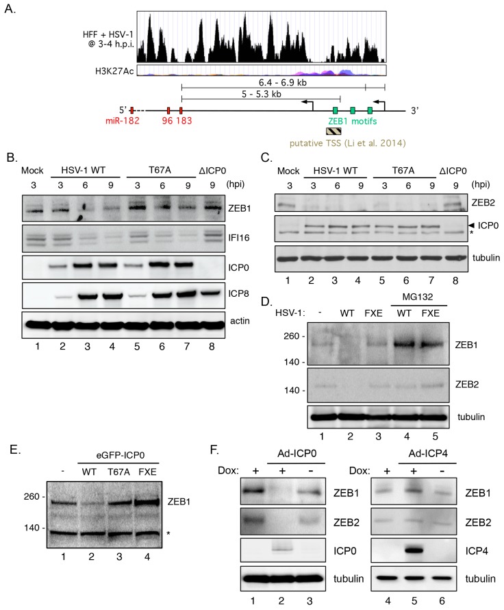
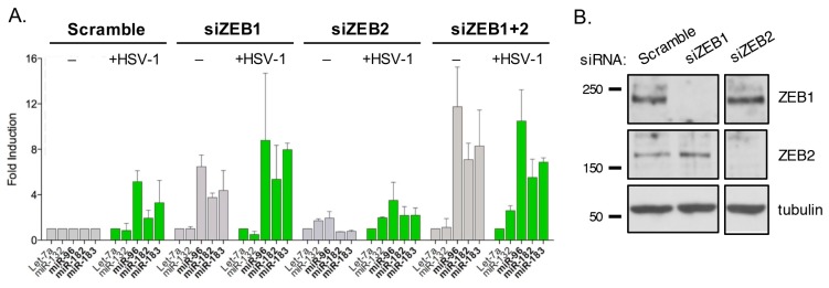
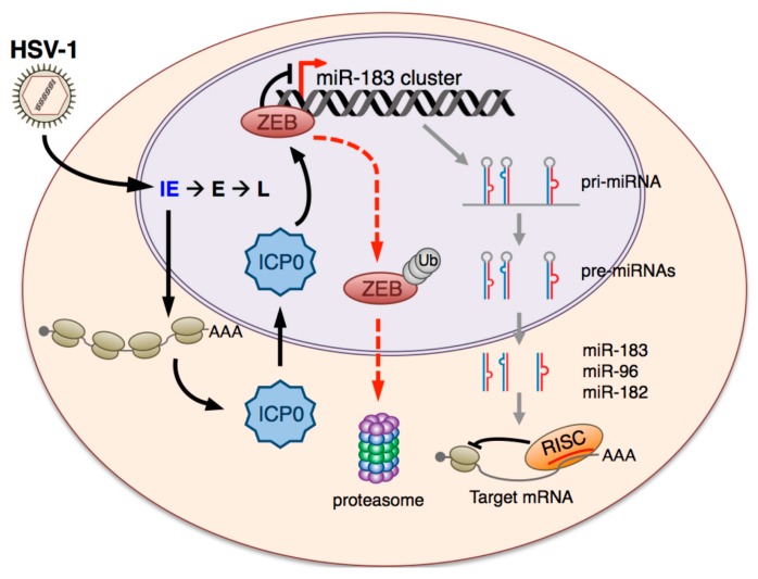
Infection with herpes simplex virus-1 (HSV-1) brings numerous changes in cellular gene expression. Levels of most host mRNAs are reduced, limiting synthesis of host proteins, especially those involved in antiviral defenses. The impact of HSV-1 on host microRNAs (miRNAs), an extensive network of short non-coding RNAs that regulate mRNA stability/translation, remains largely unexplored. Here we show that transcription of the miR-183 cluster (miR-183, miR-96, and miR-182) is selectively induced by HSV-1 during productive infection of primary fibroblasts and neurons. ICP0, a viral E3 ubiquitin ligase expressed as an immediate-early protein, is both necessary and sufficient for this induction. Nuclear exclusion of ICP0 or removal of the RING (really interesting new gene) finger domain that is required for E3 ligase activity prevents induction. ICP0 promotes the degradation of numerous host proteins and for the most part, the downstream consequences are unknown. Induction of the miR-183 cluster can be mimicked by depletion of host transcriptional repressors zinc finger E-box binding homeobox 1 (ZEB1)/-crystallin enhancer binding factor 1 (δEF1) and zinc finger E-box binding homeobox 2 (ZEB2)/Smad-interacting protein 1 (SIP1), which we establish as new substrates for ICP0-mediated degradation. Thus, HSV-1 selectively stimulates expression of the miR-183 cluster by ICP0-mediated degradation of ZEB transcriptional repressors.

Keywords: E3 ubiquitin ligase; HSV-1; ICP0; ZEB; herpes simplex virus; host shutoff; miR-182; miR-183; miR-96; microRNA.


Conflict of interest statement

The authors declare no conflict of interest. The funding sponsors had no role in the design of the study; in the collection, analyses, or interpretation of data; in the writing of the manuscript, or in the decision to publish the results.

Figures

Figure 1
Figure 1
Induction of the host miR-183/96/182 cluster during herpes simplex virus 1 (HSV-1) infection of primary fibroblasts and neurons. (A) Scatter plot showing the ratio of normalized counts for individual host miRNAs from rat superior cervical ganglia (SCG)-derived neurons that were either mock infected or infected with wild type HSV-1 GFP-Us11 (strain Patton) at a multiplicity of infection (MOI) of 1 plaque forming unit (pfu) per neuron in the presence of acyclovir [42,43,44]. Under these conditions wild type HSV-1 establishes a quiescent infection within 5–7 days, wherein the viral genome is retained in up to half of the neurons but no infectious virus can be detected [43]. Seven days after infection, total RNA was collected and used to generate small-RNA libraries that were queried by deep sequencing. Data points corresponding to miR-182 and miR-183 are highlighted; (B) Comparison of individual host miRNA counts in a quiescent infection model using growth arrested human primary fibroblasts infected with HSV-1 wild type (WT) or mock-infected; (C) Schematic showing the long arm of human chromosome 7 and organization of the microRNA miR-183/96/182 gene (miR-183C) and flanking protein coding genes, nuclear respiratory factor 1 (NRF1) and ubiquitin-conjugating enzyme E2H (UBE2H). Arrow indicates transcriptional orientation; (D) Alignment of mature miR-96-5p, miR-182-5p, and miR-183-5p sequences from human, rat, and mouse. Shading indicates the predicted seed sequences. Invariant residues are indicated with an asterisk. Residues conserved in two of the three paralogs are indicated with a diamond. Both miR-96 and miR-183 are identical in each of three species, whereas human miR-182 differs slightly at the 3′-end from the rodent counterparts.
Figure 2
Figure 2
Levels of all three members of the miR-183 cluster are induced by HSV-1 infection of primary neurons and fibroblasts. (A) Time course analysis of mature Let-7a, miR-132, miR-96, miR-182, and miR-183 levels in rat SCG-derived neuron cultures pretreated with acyclovir (ACV) for one day (at day 6 in vitro) and then infected with HSV-1 wild type (GFP-Us11 strain Patton) at an MOI of 1 pfu/neuron in the presence of ACV. RNA was harvested at days 0, 1, 2, 3, and 7 post-infection and queried by SYBR green-based reverse transcription quantitative polymerase chain reaction (RT-qPCR), with values normalized to Let-7a; (B) RT-qPCR analysis of miR-132, miR-96, miR-182, and miR-183 levels in RNA isolated from normal human dermal fibroblasts (NHDFs) infected with HSV-1 wild type (MOI = 3) relative to mock infected controls. Samples were collected at 3, 6, or 9 h post-infection (hpi). Values are normalized to Let-7a and plotted as the mean and standard error of three biological replicates; (C) Raw Ct (cycle threshold) values for the data shown in panel B, verifying the selective induction of the miR-183 cluster miRNAs; (D,E) Analysis of host miRNA abundance in HeLa (D) and U2OS (E) cells after infection with HSV-1 wild type (MOI = 3) relative to mock-infected cells. Samples were collected at 6 h post infection.
Figure 3
Figure 3
Transcription of the miR-183 cluster is induced during HSV-1 infection of primary fibroblasts. UCSC Genome Browser view of the human miR-183/96/182 cluster locus (negative strand, chromosome 7q32.2) and flanking region including the ubiquitin-conjugating enzyme E2H (UBE2H) gene showing the locations of 4sU-labeled RNA-seq reads obtained from primary human foreskin fibroblasts (HFFs) either uninfected or infected with wild type HSV-1 (strain 17) and harvested at various time points. Adapted from data (GEO database accession No. GSE59717) generated by Rutkowski, Dölken, and colleagues [27].
Figure 4
Figure 4
Induction of the host miR-183/96/182 cluster requires viral gene transcription and protein synthesis of an immediate-early viral factor. (A) Schematic representation of the HSV-1 productive (lytic) cycle gene expression cascade to illustrate the points of interruption caused by disruptive mutations in specific viral genes (ΔICP0, ΔICP4, ΔICP27, or ΔDNA polymerase) or exposure to ultraviolet (UV) radiation or protein synthesis inhibitor cycloheximide (CHX). Dashed arrows indicate reduced expression; (BG) Profiling of cluster induction in NHDFs upon infection with (B) wild type or UV-irradiated HSV-1 wild type (GFP-Us11 strain Patton); (C) HSV-1 wild type in the presence or absence of CHX; (D) a ΔDNA polymerase (early) viral mutant relative to wild type HSV-1, and (EG) immediate-early viral mutants (ΔICP0, ΔICP4, ΔICP27) relative to wild type HSV-1.
Figure 5
Figure 5
The immediate early viral factor ICP0 is sufficient to cause induction of the host miR-183/96/182 cluster. (A,B) Profiling of cluster induction relative to mock treated NHDFs, of adenoviral vectors delivering ICP0 (Ad-ICP0), ICP4 (Ad-ICP4), or VP16 (Ad-VP16) as a negative control. Lysates were prepared 48 h after infection. Statistical significance was calculated using a two-way analysis of variance (ANOVA) test, with * indicating a p-value of ≤ 0.05 and ** a p-value of ≤ 0.005, *** indicating a p-value of ≤ 0.0005, ns indicates not significant; (C) Immunoblot confirming expression of recombinant ICP0 (lane 3), ICP4 (lane 4), and VP16 (lane 5) from the adenoviral vectors. Infection with wild type HSV-1 serves a positive control (lane 2). ICP0 bands indicated by arrowhead, asterisk denotes a non-specific band. Mock (lane 1) and wild type HSV-1 (HSV-1 WT, lane 2) samples were collected 6 h post-infection, while all adenoviral samples were collected 48 h post infection; (D) Subcellular localization of ICP0 detected by indirect immunofluorescence in mock, wild type HSV-1, or ΔICP4 (n12) infections of NHDFs. Nuclei were visualized by 4′,6-diamidino-2-phenylindole (DAPI) staining; (E) Total protein was collected from mock, wild type HSV-1, and ΔICP4 (n12) infected NHDFs at 10 h post-infection and probed by immunoblotting with antibodies against viral immediate early proteins ICP4, ICP0, ICP27, and loading control, α-tubulin.
Figure 6
Figure 6
Induction of the host miR-183/96/182 cluster requires ICP0 nuclear localization and E3 ligase function to direct degradation of a host factor. (A) Schematic showing functional domains within wild type (WT) and mutant versions of HSV-1 ICP0. The RING (really interesting new gene) domain confers E3 ligase function to ICP0, and is deleted in the FXE mutant while the D8 mutant lacks an nuclear localization signal (NLS); (B) Profiling of cluster induction by WT HSV-1, FXE, and D8 virus infection of NHDFs; (C) Immunoblot detection of ICP0 expression by WT HSV-1 and respective mutant viruses. Asterisk indicates a non-specific band; (D) Schematic of ICP0 showing the location of phosphorylation residue threonine 67 and its substitution to alanine in the mutant form of ICP0 produced by the T67A virus; (E) Profiling of cluster induction by WT HSV-1 and T67A virus infection of NHDFs; (F) Immunoblot detection of ICP0 expression by WT and T67A HSV-1.
Figure 7
Figure 7
Induction of the host miR-183/96/182 cluster correlates with the degradation of host zinc finger E-box homeodomain (ZEB) transcriptional repressors by ICP0. (A) UCSC Genome Browser data from Rutkowski and colleagues [27] showing RNA-seq reads from human foreskin fibroblasts (HFFs) infected with wild type HSV-1 (HSV-1 WT) that map to the human miR-183/96/182 cluster locus and region upstream of the cluster. ENCODE data for the corresponding regions shows a region enriched for acetylated histone H3 (H3K27Ac). These regions are near sites posited to possess ZEB1 binding motifs as well as near a putative miR-183/96/182 cluster, transcription start site (TSS), as reported [53]; (B) Time course analysis of NHDFs infected with HSV-1 wild type (lanes 2–4) or T67A (lanes 5–7). Lysates were prepared at 3, 6, and 9 h post-infection. As controls, cells were either mock infected (lane 1) or infected with HSV-1 ΔICP0 (lane 8). Blotting was performed with antibodies against ZEB1, IFI16, ICP0, ICP8, and actin. Asterisk indicates a non-specific band; (C) Similar experimental setup as in B; blotting was performed using antibodies against ZEB2, ICP0, and tubulin. A non-specific band detected by the ICP0 antibody is marked with an asterisk; (D) Analysis of ZEB1 and ZEB2 levels in HFF cells either mock infected or infected with HSV-1 wild type (lanes 2 and 4) or FXE (lanes 3 and 5). For lanes 4 and 5, the proteasome inhibitor MG132 was added to the culture media prior to harvest at 8 h post infection; (E) Immunoblotting of ZEB1 in 293T cells transfected with plasmids expressing the wild type, T67A, and FXE forms of ICP0. Asterisk indicates a non-specific band; (F) HFF cells were infected with adenoviral vectors encoding ICP0 (Ad-ICP0, lanes 2 and 3) or ICP4 (Ad-ICP4, lanes 5 and 6) together with Ad.C-rtTA, an adenovirus expressing the doxycycline regulated transactivator. After infection, doxycycline was either added (+) or omitted (−) from the media.
Figure 8
Figure 8
ZEB factors regulate miR-183/96/182 cluster expression. (A) Profiling of Let-7a, miR-132, miR-96, miR-182, and miR-183 levels in NHDFs either mock (gray bars) or wild type HSV-1 (green bars) infected after transfection with non-specific (Scramble) and specific (ZEB1, ZEB2) siRNAs. The means and standard error are shown. (B) Immunoblot to confirm knockdown of ZEB1 and ZEB2 in NHDFs transfected with anti-ZEB small interfering RNAs (siRNAs).
Figure 9
Figure 9
Proposed mechanism for upregulation of the miR-183/96/182 cluster by HSV-1 in non-transformed, terminally differentiated cells such as primary fibroblasts and neurons. Prior to infection, the miR-183/96/182 locus is transcriptionally repressed by the ZEB1 and ZEB2 transcription factors. Upon infection by HSV-1, the immediate early protein ICP0 is synthesized and imported into the nucleus, where it acts as an E3 ubiquitin-ligase to promote degradation of the ZEB proteins by the proteasome. This leads to de-repression of the transcriptionally silenced miR-183/96/182 locus and increased synthesis of the pri-miRNA transcript, which is subsequently exported and processed to produce the mature miR-183, miR-96, and miR-182 miRNA, which may become functional after incorporation into the RNA-induced silencing complex (RISC).

Similar articles

Cited by

References

    1. Bartel D.P. MicroRNAs: Genomics, biogenesis, mechanism, and function. Cell. 2004;116:281–297. doi: 10.1016/S0092-8674(04)00045-5. - DOI - PubMed
    1. Bartel D.P. MicroRNAs: Target recognition and regulatory functions. Cell. 2009;136:215–233. doi: 10.1016/j.cell.2009.01.002. - DOI - PMC - PubMed
    1. O’Connor C.M., Vanícek J., Murphy E.A. Host microRNA regulation of human cytomegalovirus immediate early protein translation promotes viral latency. J. Virol. 2014;88:5524–5532. doi: 10.1128/JVI.00481-14. - DOI - PMC - PubMed
    1. Powdrill M.H., Desrochers G.F., Singaravelu R., Pezacki J.P. The role of microRNAs in metabolic interactions between viruses and their hosts. Curr. Opin. Virol. 2016;19:71–76. doi: 10.1016/j.coviro.2016.07.005. - DOI - PubMed
    1. Chen L., Song Y., He L., Wan X., Lai L., Dai F., Liu Y., Wang Q. MicroRNA-223 promotes type I interferon production in antiviral innate immunity by targeting forkhead box protein O3 (FOXO3) J. Biol. Chem. 2016;291:14706–14716. doi: 10.1074/jbc.M115.700252. - DOI - PMC - PubMed

Publication types

MeSH terms

LinkOut - more resources