. 2017 Nov 23;36(47):6568-6580.
doi: 10.1038/onc.2017.248. Epub 2017 Aug 7.

Glioblastoma stem cells exploit the αvβ8 integrin-TGFβ1 signaling axis to drive tumor initiation and progression

Affiliations

Glioblastoma stem cells exploit the αvβ8 integrin-TGFβ1 signaling axis to drive tumor initiation and progression

P A Guerrero et al. Oncogene. .

Abstract

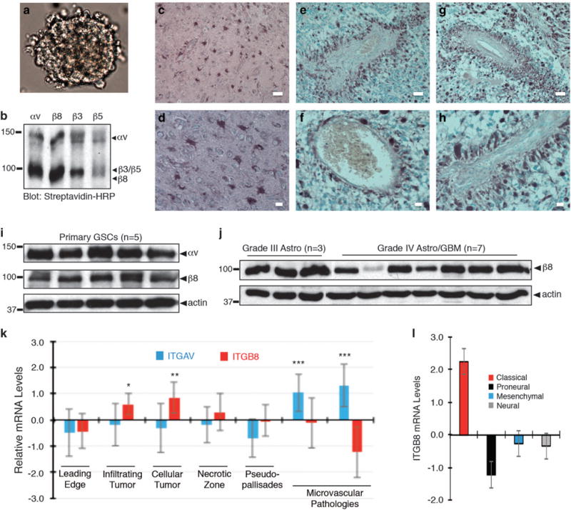
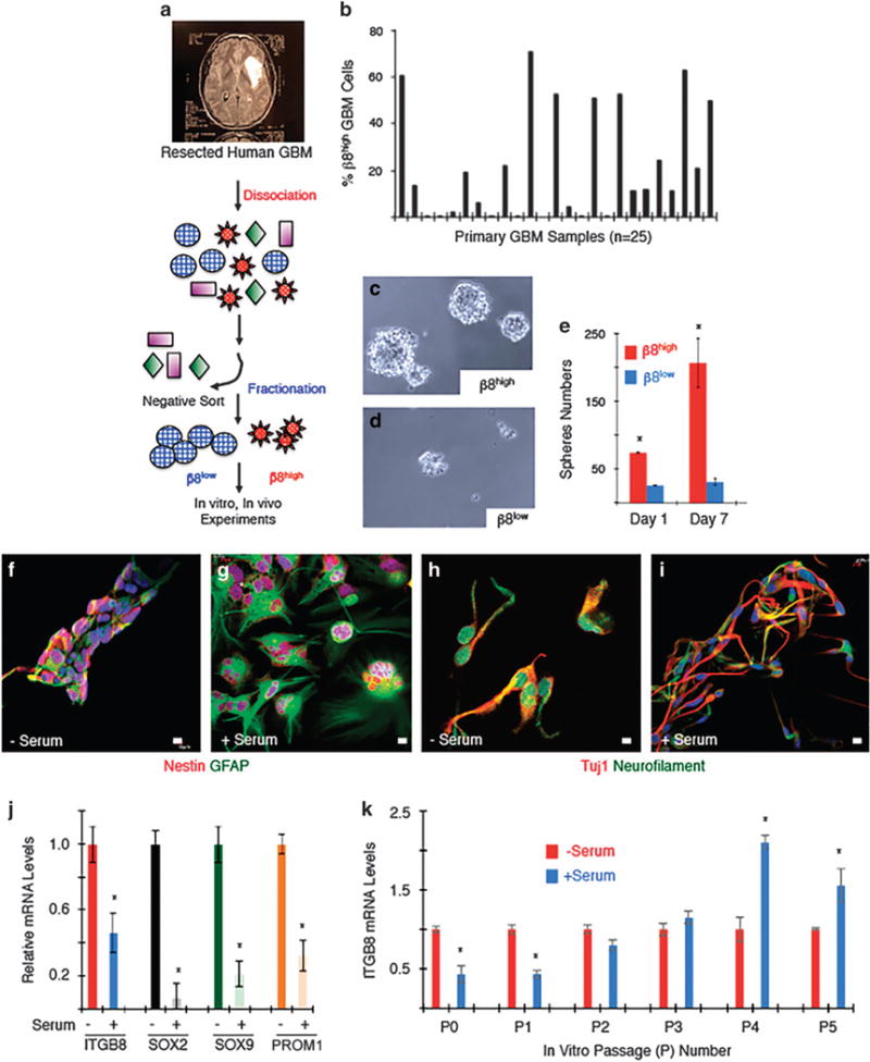
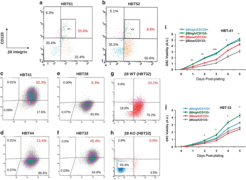
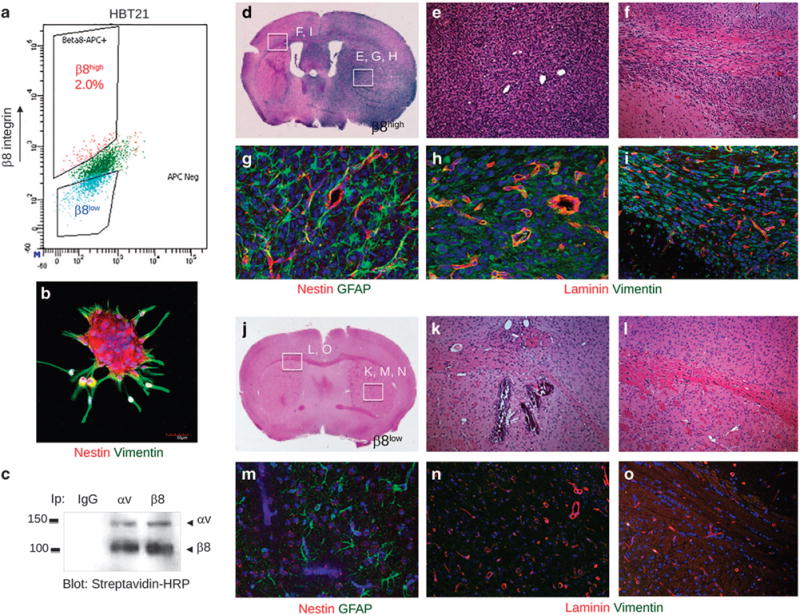
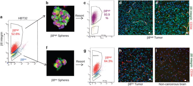
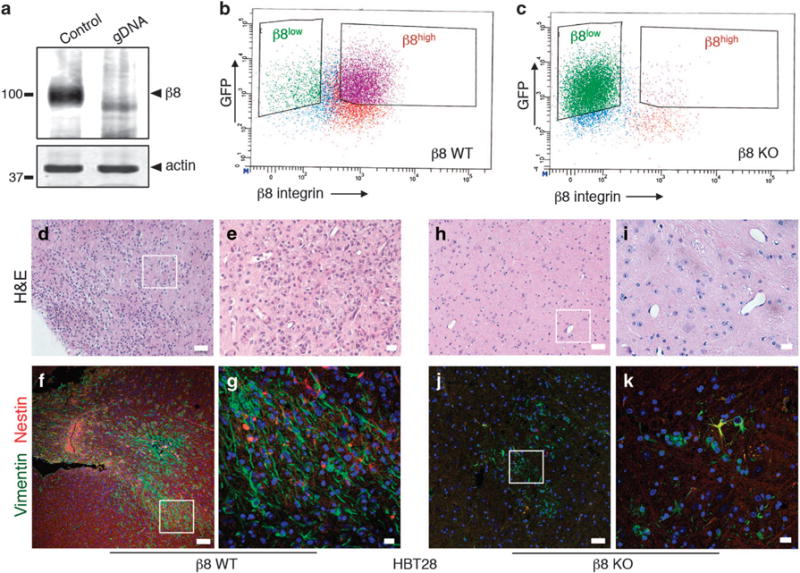
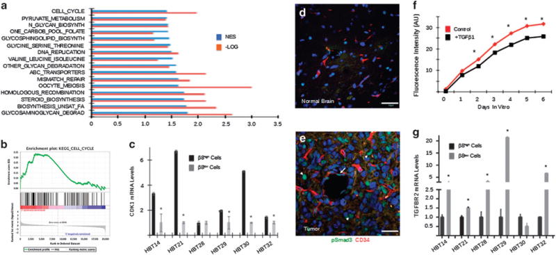
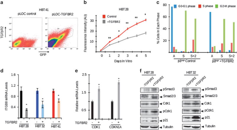
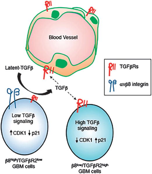
Glioblastoma (GBM) is a primary brain cancer that contains populations of stem-like cancer cells (GSCs) that home to specialized perivascular niches. GSC interactions with their niche influence self-renewal, differentiation and drug resistance, although the pathways underlying these events remain largely unknown. Here, we report that the integrin αvβ8 and its latent transforming growth factor β1 (TGFβ1) protein ligand have central roles in promoting niche co-option and GBM initiation. αvβ8 integrin is highly expressed in GSCs and is essential for self-renewal and lineage commitment in vitro. Fractionation of β8high cells from freshly resected human GBM samples also reveals a requirement for this integrin in tumorigenesis in vivo. Whole-transcriptome sequencing reveals that αvβ8 integrin regulates tumor development, in part, by driving TGFβ1-induced DNA replication and mitotic checkpoint progression. Collectively, these data identify the αvβ8 integrin-TGFβ1 signaling axis as crucial for exploitation of the perivascular niche and identify potential therapeutic targets for inhibiting tumor growth and progression in patients with GBM.


Conflict of interest statement

CONFLICT OF INTEREST

The authors declare no conflict of interest.

Figures

Figure 1
Figure 1
β8 integrin is expressed in cultured GBM spheroids and is enriched in perivascular GBM cells in situ. (a) Primary tumor cells cultured from freshly resected GBM tissue grow as neurosphere-like spheroids in serum-free media containing EGF and bFGF. (b) Cell surface biotinylation and immunoprecipitation experiments identify αvβ8 as a major αv integrin-containing heterodimeric protein expressed in primary human GBM cells. (c–h) Immunohistochemistry staining with an anti-β8 integrin antibody reveals integrin protein expression patterns in non-cancerous brain and human GBM samples. Note that β8 integrin is expressed in reactive astrocytes in the non-cancerous brain (c, d). In contrast, within brain tumors β8 integrin protein is enriched in perivascular GBM cells (e–h). Scale bar, 20 μm. (i); Immunoblot analysis of αv and β8 integrin proteins in human GBM spheroids (n = 5). (j) Immunoblot analysis of β8 integrin protein levels in different tumor lysates from grade III astrocytomas (n = 3) and grade IV GBM lysates (n = 7). (k) Differential expression of ITGAV and ITGB8 mRNAs in various tumor regions based on querying the IVY GBM Atlas Project. (l) Analysis of the TCGA GBM database identifies ITGB8 as a molecular marker for the classical GBM sub-type, *P<0.05, **P<0.01, ***P<0.001.
Figure 2
Figure 2
β8 integrin is required for GSC self-renewal and differentiation in vitro. (a) Experimental approach for isolation of β8high and β8low primary GBM cells from human samples followed by in vitro culturing and/or intracranial injection. (b) Summary of β8 integrin protein expression levels as determined by FACS in 25 different freshly resected primary GBM samples. (c, d) β8high GBM cells from sample HBT14 form spheroids and survive in culture (c), whereas β8low cells do not form spheroids and fail to thrive in culture (d). Images shown are of spheroids formed from non-passaged β8high and β8low GBM cells. (e) Quantitation of β8 integrin-dependent sphere formation in vitro. (f–i) Spheroids generated from low-passage β8high GBM cells (HBT41) were grown in the presence or absence of serum and immunofluorescently labeled with anti-Nestin and anti-GFAP to label neural stem cells and astrocytes (f, g) or anti-TUJ1 and anti-neurofilament antibodies to label neurons (h, i). (j) Neural stem cell markers were quantified by RT-PCR using spheroids generated from low-passage β8high GBM cells (HBT28) before and after differentiation via serum exposure. (k) Serum-induced differentiation of β8high GBM cells (HBT32) leads to reduced β8 integrin expression at low passages, but β8 integrin expression increases in more differentiated cells at higher passages.
Figure 3
Figure 3
Analysis of β8 integrin and CD133 protein expression in primary GSCs. (a, b) Primary tumor cells from two different freshly resected GBM samples were analyzed for β8 integrin and CD133 expression. Note that the majority of β8high GBM cells do not express CD133. (c–f) Unfractionated primary tumor cells (c, d) from two different freshly resected GBM samples (HBT41 and HBT44) were cultured in serum-free media. Alternatively, β8high GBM cells were fractionated (HBT28 and HBT32) and cultured in vitro (e, f). Note that nearly all GBM cells, whether sorted for β8 integrin or not, express high levels of β8 integrin protein. CD133 protein levels are more variable and do not fully coincide with β8 integrin expression. (g, h) Crispr-Cas9 strategies were used to target ITGB8 in spheroids formed from β8high GBM cells (HBT28) followed by FACS analysis. Note that CD133 is absent following ITGB8 gene targeting. Validation of ITGB8 gene editing via Crispr-Cas9 and absence of integrin protein expression is detailed in Figure 6 and Supplementary Figure 10. (i, j) GBM cells from HBT41 (i) and HBT32 samples (j) were fractionated by FACS based on differential expression of CD133 and β8 integrin. Cell growth and viability were quantified in spheroids every day for 5 days. In comparison with β8high/CD133 cells, note that β8low/CD133+ and β8low/CD133 cell fractions show reduced viability, *P<0.05, **P<0.01, ***P<0.001. β8high/CD133+.
Figure 4
Figure 4
β8 integrin is essential for GBM initiation, growth and invasion in vivo. (a) Representative FACS plot of β8high and β8low primary GBM cell fractions from a freshly resected tumor sample (HBT21). (b) Spheroids formed from sorted β8high cells were labeled with anti-Nestin (red) and anti-Vimentin (green) antibodies. (c) αvβ8 integrin heterodimeric protein is robustly expressed in spheroids formed from sorted β8high cells, as revealed by cell surface biotinylation and co-immunoprecipitation. (d–f) Images of H&E-stained brain sections from mice injected with β8high GBM cells sorted from sample HBT21. Note that β8high cells form diffuse, intracranial tumors that invade along white matter tracts and blood vessels. (g–i) Immunofluorescence analysis of β8 integrin-dependent GBM growth, invasion and angiogenesis in xenograft tumors. β8high GBM cells generated well-vascularized and invasive tumors as revealed by anti-laminin staining to identify vascular basement membranes and anti-vimentin staining to identify human cells. (j–l) Images of H&E-stained tumor sections from mice injected with β8low GBM cells from the HBT21 sample. (m–o) Immunofluorescence analysis of mice injected with β8low GBM cells, revealing absence of vimentin-expressing human tumor cells.
Figure 5
Figure 5
β8 integrin is re-expressed in spheroids and tumors generated from β8low GBM cells. (a) FACS-based fractionation of β8high and β8low tumor cells from a freshly resected primary GBM sample (HBT32). (b, c) β8high GBM cells form spheroids in culture (b) that express the neural stem cell marker Nestin (red) and the more differentiated cell marker Vimentin (green). FACS analysis of spheroids reveals β8 integrin protein expression in nearly all primary GBM cells (c). (d, e) β8high GBM cells fractionated from HBT32 (a) were intracranially implanted into the mouse brain (n = 3 mice injected per cell type). β8high cells formed large and invasive brain tumors in mice and express β8 integrin protein, as revealed with a human-specific anti-β8 integrin protein. Note that perivascular tumor cells express robust levels of β8 integrin protein. (f, g) β8low GBM cells from HBT32 form spheroids in culture (f) that express Nestin (red) and Vimentin (green), although they are smaller than spheroids formed from β8high GBM cells. FACS analysis of β8low spheroids reveals β8 integrin protein expression in 64% of GBM cells (g). (h) Immunofluorescence analysis reveals upregulated integrin protein expression tumors generated from β8low GBM cells. (i) A non-cancerous region of the mouse brain was used to control for specificity of the human-specific β8 integrin antibody. All scale bars are 10 μm.
Figure 6
Figure 6
Genetically targeting β8 integrin in low-passage GBM cells leads to defective tumor initiation in vivo. (a) Immunoblot analysis showing absence of β8 integrin protein expression in β8KO cells generated via Crispr-Cas9 gene editing strategies. (b, c) FACS plot from β8WT (b) and β8KO (c) GBM cells, revealing loss of β8 integrin protein expression on the GBM cell surface. (d, e) Images of H&E-stained tumor sections from mice injected with β8WT GBM cells, revealing larger and more invasive tumors derived from β8WT cells. (f, g) Immunofluorescence analysis of xenograft tumors derived from β8WT GBM cells. The anti-vimentin antibody (green) is specific for human cells and anti-nestin (red) labels neural stem cells. (h, i) Brain sections from mice injected with β8KO GBM cells were stained with H&E, revealing small and minimally invasive tumors. (j, k) Immunofluorescence analysis with anti-vimentin and anti-nestin antibodies of mouse brains injected with β8KO GBM cells. Note that there are fewer human cells in β8KO-derived tumors as revealed by anti-vimentin immunofluorescence staining. All scale bars are 10 μm.
Figure 7
Figure 7
Analysis of β8 integrin-dependent gene expression signatures in GSCs. (a) β8high and β8low GBM cells were fractionated from three different freshly resected primary human tumor samples and analyzed by whole-transcriptome sequencing. Multiple signaling pathways were identified by gene set enrichment analysis based on differential β8 integrin expression. The normalized enrichment score (NES) and the log transformed (−Log) P-values are shown for the top 16 pathways. (b) NES pathway analysis of β8 integrin-dependent cell cycle gene expression signatures in sorted β8high GBM cells. (c) RT-PCR validation showing that CDK1 mRNA expression is significantly downregulated in β8low GBM cells fractionated from five of six human tumor samples. (d, e) Sections from normal mouse brain (d) or from xenograft tumors formed from β8high GBM cells (e) were immunofluorescently labeled with anti-pSmad3 and anti-CD34. Note that TGFβ receptor signaling is active within intratumoral blood vessels (arrow) and in tumor cells (arrowheads). All scale bars are 50 μm. (f) in vitro growth analysis of β8high GBM cells treated with TGFβ1, revealing TGFβ1-dependent growth suppression. (g) RT-PCR data with five different GBM samples showing elevated levels of TGFBR2 mRNA in β8low GBM cells versus β8high GBM cells.
Figure 8
Figure 8
The αvβ8 integrin-TGFβ1 signaling pathway regulates mitotic checkpoint progression in primary GBM cells. (a) GBM cells expressing control pLOC lentivirus (left) or pLOC containing a TGFBR2 cDNA (right) were isolated by FACS based on expression of TGFβR2. (b) β8high GBM cells forcibly expressing TGFβR2 show diminished growth in vitro. (c) Cell cycle phases were analyzed in cultured β8high GBM cells that were asynchronous (A), synchronized (S), or released from synchronization for 2 hours (S+2). Cells were previously infected with control pLOC lentivirus or pLOC expressing TGFβR2. Note that in comparison to controls, TGFβR2 expression results in diminished percentages of cells in the G2-M phase. (d) β8high GBM cells infected with control lentivirus or lentiviruses expressing TGFβR2 were analyzed by RT-PCR, revealing diminished levels of ITGB8 mRNA. (e) β8high GBM cells infected with control lentivirus or lentivirus expressing TGFβR2 were analyzed by RT-PCR, revealing TGFβ1-dependent increases in CDK1 and CDKN1A mRNAs. (f) β8high GBM cells fractionated from two different tumors were infected with pLOC or pLOC-TGFβR2 lentivirus. Detergent-soluble lysates were analyzed by immunoblotting. Note that forced TGFβR2 expression leads to increased levels of phosphorylated Smad3 and Cdk1 proteins as well as elevated expression of p21cip protein.
Figure 9
Figure 9
A model for the αvβ8 integrin-TGFβ1 signaling axis in the GBM perivascular niche. β8high GBM cells mediate adhesion to latent-TGFβ1/3 in the ECM, leading to activation of TGFβ receptor signaling in β8low GBM cells and/or in vascular endothelial cells. Varying degrees of TGFβ receptor signaling in β8low versus β8high GBM cells impacts cell cycle progression via CDK1 and CDKN1A/p21cip, thus impacting levels of their proliferation and/or differentiation.

Similar articles

Cited by

References

    1. Alcantara Llaguno S, Chen J, Kwon CH, Jackson EL, Li Y, Burns DK, et al. Malignant astrocytomas originate from neural stem/progenitor cells in a somatic tumor suppressor mouse model. Cancer cell. 2009;15:45–56. - PMC - PubMed
    1. Zheng H, Ying H, Yan H, Kimmelman AC, Hiller DJ, Chen AJ, et al. p53 and Pten control neural and glioma stem/progenitor cell renewal and differentiation. Nature. 2008;455:1129–1133. - PMC - PubMed
    1. Lathia JD, Mack SC, Mulkearns-Hubert EE, Valentim CL, Rich JN. Cancer stem cells in glioblastoma. Genes Dev. 2015;29:1203–1217. - PMC - PubMed
    1. Sanai N, Alvarez-Buylla A, Berger MS. Neural stem cells and the origin of gliomas. N Engl J Med. 2005;353:811–822. - PubMed
    1. Bao S, Wu Q, McLendon RE, Hao Y, Shi Q, Hjelmeland AB, et al. Glioma stem cells promote radioresistance by preferential activation of the DNA damage response. Nature. 2006;444:756–760. - PubMed

Publication types

MeSH terms