. 2017 Sep 5;56(35):4626-4636.
doi: 10.1021/acs.biochem.7b00618. Epub 2017 Aug 23.

A Noncanonical Binding Site in the EVH1 Domain of Vasodilator-Stimulated Phosphoprotein Regulates Its Interactions with the Proline Rich Region of Zyxin

Affiliations

A Noncanonical Binding Site in the EVH1 Domain of Vasodilator-Stimulated Phosphoprotein Regulates Its Interactions with the Proline Rich Region of Zyxin

Lucila Andrea Acevedo et al. Biochemistry. .

Abstract

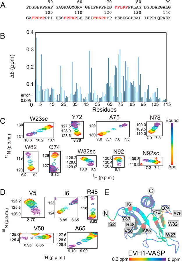
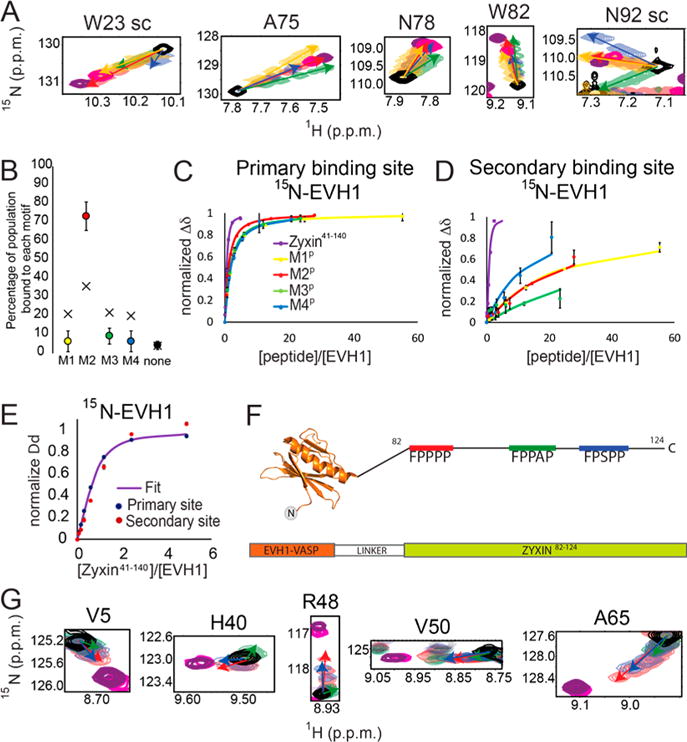
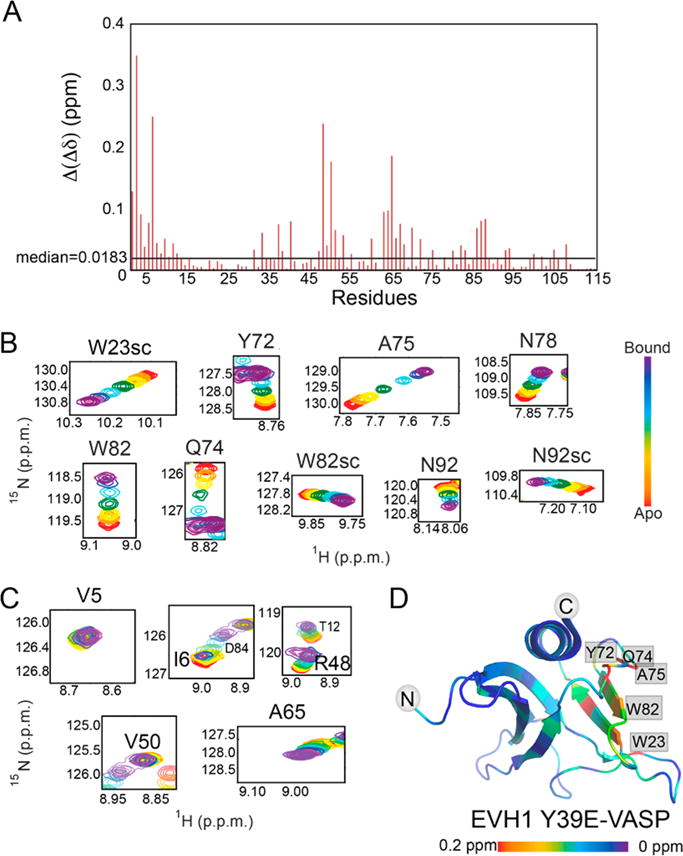
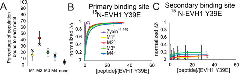
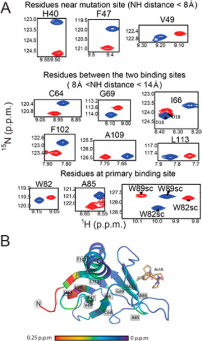
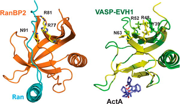
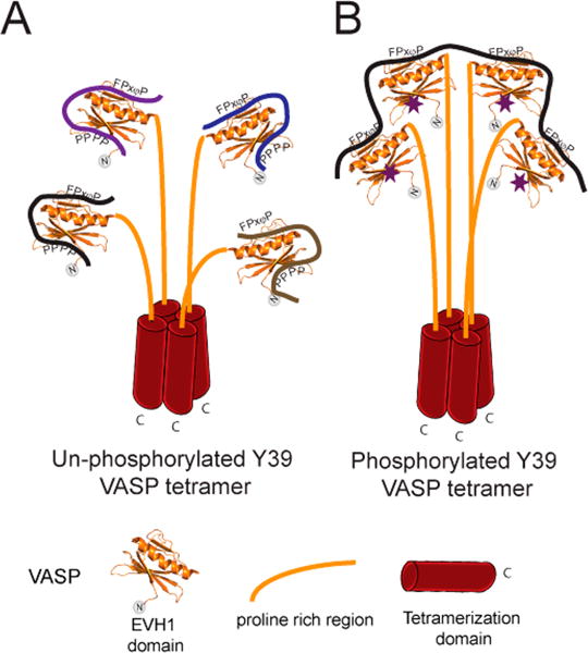
Vasodilator-stimulated phosphoprotein (VASP) is a processive actin polymerase with roles in the control of cell shape and cell migration. Through interaction with the cytoskeletal adaptor protein Zyxin, VASP can localize to damaged stress fibers where it serves to repair and reinforce these structures. VASP localization is mediated by its N-terminal Ena/VASP homology (EVH1) domain, which binds to the (W/F)PxφP motif (most commonly occurring as FPPPP) found in cytoskeletal proteins such as vinculin, lamellipodin, and Zyxin. Sequentially close clusters of four or five of these motifs frequently occur, as in the proline rich region of Zyxin with four such motifs. This suggests that tetrameric VASP might bind very tightly to Zyxin through avidity, with all four EVH1 domains binding to a single Zyxin molecule. Here, quantitative nuclear magnetic resonance titration analysis reveals a dominant bivalent 1:1 (Zyxin:EVH1) interaction between the Zyxin proline rich region and the VASP EVH1 domain that utilizes the EVH1 canonical binding site and a novel secondary binding site on the opposite face of the EVH1 domain. We further show that binding to the secondary binding site is specifically inhibited by mutation of VASP EVH1 domain residue Y39 to E, which mimics Abl-induced phosphorylation of Y39. On the basis of these findings, we propose a model in which phosphorylation of Y39 acts as a stoichiometry switch that governs binding partner selection by the constitutive VASP tetramer. These results have broader implications for other multivalent VASP EVH1 domain binding partners and for furthering our understanding of the role of Y39 phosphorylation in regulating VASP localization and cellular function.


Conflict of interest statement

Notes

Theauthors declare no competing financial interest.

Figures

Figure 1
Figure 1
Zyxin41–140 titration provides evidence of a secondary binding site in the EVH1 domain of VASP. (A) Amino acid sequence of the Zyxin41–140 construct, highlighting the four VASP binding motifs (red, bold type). (B) Composite chemical shift changes in [15N]EVH1 induced by addition of Zyxin41–140, calculated using the equation Δδ=(0.154ΔδN)2+ΔδH 2. Extracted regions of overlays of 15N–1H HSQC spectra of [15N]EVH1 titrated with Zyxin41–140 show trajectories for residues in (C) the canonical binding site and (D) a novel secondary site, all of which show maximum Δδ values in excess of 10 times the error in Δδ (determined to be 0.005). (E) Chemical shift changes mapped onto the structure of VASP EVH1 (Protein Data Bank entry 1EGX) show that Zyxin41–140 induces chemical shift perturbations for residues in the primary binding site (far right) but also for residues on the opposite face of the EVH1 domain (far left). Red denotes the largest chemical shift perturbation and purple no perturbation.
Figure 2
Figure 2
Secondary binding site that creates a larger population of motif 2 bound to the canonical binding site. (A) Titrations show distinct chemical shift peak trajectories upon addition of each motif peptide (yellow, M1P; red, M2P; green, M3P; blue, M4P). The magenta peak shows titration with Zyxin41–140 at the highest Zyxin concentration. Purple shows the chemical shift of chimeric EVH1-Zyxin. (B) Distribution of Zyxin41–140 motifs bound at the canonical site of EVH1 deduced from chemical shifts (filled circles) compared with the expected distribution based on Kd values of individual peptides (stars). (C) Binding curves from average normalized chemical shift changes of 10 residues in the canonical binding site of EVH1 for individual peptide titrations and for Zyxin41–140. The yellow binding curve indicates titration with M1P, the red binding curve titration of EVH1 with M2P, the green binding curve titration of EVH1 with M3P, the blue binding curve titration of EVH1 with M4P, and the purple binding curve titration of EVH1 with Zyxin41–140. (D) Like panel C, for six residues at the novel secondary binding site of EVH1. (E) Binding curve from average normalized chemical shift changes for the primary binding site (blue dots) and for the secondary binding site (red dots) and a global fit to both curves (Kd = 32.2 μM). (F) Chimera of EVH1 and Zyxin (EVH1–Zyxin82–124) containing M2 (red), M3 (green), and M4 (blue) motifs. (G) Overlays of 15N–1H HSQC spectra for residues in the noncanonical site, titrated with M2P (red), M3P (green), M4P (green), or Zyxin41–140 (magenta) or in the context of EVH1–Zyxin82–124 (purple).
Figure 3
Figure 3
Phosphomimetic mutation abolishes secondary site binding. (A) Bar plot showing differences between composite chemical shift perturbations of the 1H and 15N resonances upon addition of Zyxin41–140 to EVH1 WT and EVH1 Y39E. Those residues with the largest perturbation differences are located in the novel secondary binding site. Overlays of 15N–1H HSQC spectra for residues in (B) the canonical binding site and (C) the secondary site show binding for the primary but not secondary sites. (D) Mapping chemical shift changes induced by titration of [15N]EVH1 Y39E with Zyxin41–140 onto the structure of the VASP EVH1 domain (Protein Data Bank entry 1EGX) shows large perturbations for residues in the primary binding site (far right) but almost no perturbations for residues on the opposite face of the EVH1 Y39E domain (far left). Red indicates the largest chemical shift perturbation and purple no perturbation.
Figure 4
Figure 4
Population and binding curves for [15N]EVH1 Y39E. (A) Distribution of Zyxin41–140 motifs bound at the canonical site of EVH1 Y39E deduced from chemical shifts (filled circles) compared with the expected distribution based on Kd values of individual peptides (black crosses). (B) Binding curves from average normalized chemical shift changes of 10 residues in the canonical binding site of EVH1 Y39E for individual peptide titrations and for Zyxin41–140. The yellow binding curve indicates titration with M1P, the red binding curve titration of EVH1 Y39E with M2P, the green binding curve titration of EVH1 Y39E with M3P, the blue binding curve titration of EVH1 Y39E with M4P, and the purple binding curve titration of EVH1 Y39E with Zyxin41–140. (C) Like panel B, for six residues at the novel secondary binding site of EVH1 Y39E.
Figure 5
Figure 5
1H–15N chemical shift differences between wild-type EVH1 and mutant Y39E. (A) Chemical shift differences between wild-type EVH1 (blue spectra) and EVH1 Y39E (red spectra). Both spectra were recorded under identical buffer conditions and with the same parameters. All highlighted residues display differences in chemical shift that exceed the error in chemical shift value by a factor of at least 10. (B) Mapping of 1H–15N backbone chemical shift differences to the EVH1 structure. Significant changes (>0.25 ppm) are represented by red, and no change is shown as purple.
Figure 6
Figure 6
Tyr39 is located centrally in the secondary EVH1 binding site. In the left panel, the first Ran binding domain of RanBP2 (orange) binds Ran (cyan) using a secondary binding surface similar to the site we describe, employing residues R77, R81, and N91 that correspond to R48, R52, and N63, respectively, in VASP EVH1 (Protein Data Bank entry 1RRP). In the right panel, VASP EVH1 residue Y39 is located centrally in the secondary binding site. EVH1 is colored according to chemical shift perturbations induced by Zyxin41–140 (green, no perturbation; yellow, largest perturbation). Key residues are shown as sticks. The ActA sequence FPPPP (yellow) is shown bound in the primary site.
Figure 7
Figure 7
Proposed model for the stoichiometry switch controlled by phosphorylation of Y39 in VASP tetramers. (A) In the extreme case where all Y39 residues are unphosphorylated, each EVH1 domain in the VASP tetramer could interact with a molecule of Zyxin or any other binding partner. Bivalent interactions could form due to the presence of nearby proline rich motifs. (B) In the extreme case where each Y39 residue is phosphorylated (purple stars), one molecule with multiple FPxφP motifs (like Zyxin, ActA, or lammellipodin) could bind to one tetramer of VASP. Phosphorylation of Y39 could recruit VASP exclusively to a specific binding partner with multiple canonical binding sites, while dephosphorylation could cause cross-linking between multiple binding partners.

References

    1. Breitsprecher D, Kiesewetter AK, Linkner J, Urbanke C, Resch GP, Small JV, Faix J. Clustering of VASP actively drives processive, WH2 domain-mediated actin filament elongation. EMBO J. 2008;27:2943–2954. - PMC - PubMed
    1. Breitsprecher D, Kiesewetter AK, Linkner J, Vinzenz M, Stradal TE, Small JV, Curth U, Dickinson RB, Faix J. Molecular mechanism of Ena/VASP-mediated actin-filament elongation. EMBO J. 2011;30:456–467. - PMC - PubMed
    1. Krause M, Dent EW, Bear JE, Loureiro JJ, Gertler FB. Ena/VASP proteins: regulators of the actin cytoskeleton and cell migration. Annu Rev Cell Dev Biol. 2003;19:541–564. - PubMed
    1. Bear JE, Svitkina TM, Krause M, Schafer DA, Loureiro JJ, Strasser GA, Maly IV, Chaga OY, Cooper JA, Borisy GG, Gertler FB. Antagonism between Ena/VASP proteins and actin filament capping regulates fibroblast motility. Cell. 2002;109:509–521. - PubMed
    1. Lebrand C, Dent EW, Strasser GA, Lanier LM, Krause M, Svitkina TM, Borisy GG, Gertler FB. Critical role of Ena/VASP proteins for filopodia formation in neurons and in function downstream of netrin-1. Neuron. 2004;42:37–49. - PubMed

Publication types