. 2016 Jul 29;1(1):aag0851.
doi: 10.1126/sciimmunol.aag0851. Epub 2016 Jul 29.

Immune perturbations in HIV-1-infected individuals who make broadly neutralizing antibodies

Affiliations

Immune perturbations in HIV-1-infected individuals who make broadly neutralizing antibodies

M Anthony Moody et al. Sci Immunol. .

Abstract

Induction of broadly neutralizing antibodies (bnAbs) is a goal of HIV-1 vaccine development. bnAbs occur in some HIV-1-infected individuals and frequently have characteristics of autoantibodies. We have studied cohorts of HIV-1-infected individuals who made bnAbs and compared them with those who did not do so, and determined immune traits associated with the ability to produce bnAbs. HIV-1-infected individuals with bnAbs had a higher frequency of blood autoantibodies, a lower frequency of regulatory CD4+ T cells, a higher frequency of circulating memory T follicular helper CD4+ cells, and a higher T regulatory cell level of programmed cell death-1 expression compared with HIV-1-infected individuals without bnAbs. Thus, induction of HIV-1 bnAbs may require vaccination regimens that transiently mimic immunologic perturbations in HIV-1-infected individuals.


Figures

Figure 1
Figure 1
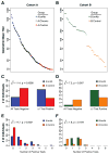
Neutralization and autoantibody testing of HIV-1-infected individuals. HIV-1 neutralization data are shown as geometric mean titer for a panel of isolates for Cohort A (A) and B (B). Individuals with the highest (bnAb) and lowest (Control) HIV-1 neutralization for each cohort were tested in autoantibody assays. Panels C and D compare the frequency of individuals in the bnAb and control groups with any positive result for Cohorts A and B, respectively. The number of positive autoantibody results is shown for Cohort A (panel E) and B (panel F).
Figure 2
Figure 2
Comparison of plasma antibody responses. When compared with the A.Control group, the A.bnAb group had higher binding to HIV-1 Env gp120 (A) and gp41 (B) but similar binding (C) to trivalent inactivated influenza vaccine for 2008 (TIV 2008). Binding was similar between the B.bnAb and B.Control groups for the same three antigens (D – F). Each symbol represents data from an individual subject; group medians, range, and quartiles are shown.
Figure 3
Figure 3
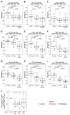
T cell subsets in Cohort A subjects with and without bnAbs and matched HIV-1-seronegative controls. Total CD4+ T cell frequency within lymphocytes was lower in Cohort A HIV-1-infected individuals vs. HIV-1-seronegative controls and was lowest in the A.bnAb group (A). Resting memory T follicular helper (mTfh) cells were elevated in Cohort A HIV-1-infected individuals vs. HIV-seronegative controls, and were highest in the A.bnAb group (B). CD4+ Treg cell frequency was lowest in the A.bnAb group (C). PD-1 MFI on CD4+ Treg cells was highest in the A.bnAb group (D). The proportion of Tfr cells within circulating CD4+ follicular-phenotype T cells in the A.bnAb group did not differ significantly from that in the A.Control or seronegative groups (E). The Tfr/Tfh ratio, defined as Tfr/Tfh=%CD25+Foxp3+cellswithinCXCR5+CD4+Tcells%non-CD25+Foxp3+cellswithinCXCR5+CD4+Tcells in the A.bnAb group did not differ significantly from that in the A.Control or seronegative groups (F). The MFI of PD-1 staining on CD4+ Tfr cells was highest in the A.bnAb group (G). Each symbol represents data from an individual subject; group medians, range, and quartiles are shown.
Figure 4
Figure 4
Phenotypic and functional analysis of CD4+ Treg cells. CD4+ Treg cells expressing HLA-DR were higher in the A.bnAb group vs. the A.Control or seronegative groups (A). Total CD4+ Treg cells expressing CTLA-4 were also higher in the A.bnAb group vs. the A.Control or seronegative groups (B); no difference between groups was found for CD4+ Treg cells expressing LAG-3 (C). In the A.bnAb group, the PD-1high subset of CD4+ Treg cells expressed higher levels of HLA-DR (D), CTLA-4 (E) and LAG-3 (F) than the PD-1low or PD-1 negative subsets of CD4+ Treg cells. The results were similar for the HIV-1 seronegative control group (G, H and I). In panels A–I, symbols represent individual subjects; group medians, range, and quartiles are shown. In experiments performed with samples from healthy HIV-1-seronegative UK donors, PD-1 negative and PD-1low CD4+ Treg cells suppressed the proliferation of conventional CD4+ T cells (Tconv) compared to that observed in the presence of other conventional CD4+ T cells, whereas PD-1high CD4+ Treg cells did not do so (J). In panel J, the symbols represent individual subjects; group medians, range and quartiles are shown; and the horizontal dashed red line indicates the level of proliferation of conventional CD4+ T cells in the presence of other conventional CD4+ T cells to which other values were normalized.
Figure 5
Figure 5
T cell subsets in healthy, HIV-1-seronegative African individuals with and without autoantibodies. HIV-1-seronegative subjects with and without autoantibodies had similar frequencies of total CD4+ T cells within lymphocytes (A). The circulating frequency of resting memory T follicular helper (mTfh) cells was similar in HIV-1-seronegative individuals with and without autoantibodies (B). CD4+ Treg cell frequency within lymphocytes in HIV-1-seronegative individuals with autoantibodies did not differ significantly from that in those without autoantibodies (C). PD-1 MFI on CD4+ Treg cells in HIV-1-seronegative individuals with autoantibodies did not differ significantly from that in those without autoantibodies (D). In all panels, each symbol represents data from an individual subject; group medians, range, and quartiles are shown.

Comment in

References

    1. Fauci AS, Marston HD. Ending AIDS--is an HIV vaccine necessary? N Engl J Med. 2014;370:495–498. - PubMed
    1. Mascola JR, Haynes BF. HIV-1 neutralizing antibodies: understanding nature’s pathways. Immunol Rev. 2013;254:225–244. - PMC - PubMed
    1. Burton DR, Mascola JR. Antibody responses to envelope glycoproteins in HIV-1 infection. Nat Immunol. 2015;16:571–576. - PMC - PubMed
    1. Rerks-Ngarm S, Pitisuttithum P, Nitayaphan S, Kaewkungwal J, Chiu J, Paris R, Premsri N, Namwat C, de Souza M, Adams E, Benenson M, Gurunathan S, Tartaglia J, McNeil JG, Francis DP, Stablein D, Birx DL, Chunsuttiwat S, Khamboonruang C, Thongcharoen P, Robb ML, Michael NL, Kunasol P, Kim JH. Vaccination with ALVAC and AIDSVAX to Prevent HIV-1 Infection in Thailand. N Engl J Med. 2009;361:2209–2220. - PubMed
    1. Montefiori DC, Karnasuta C, Huang Y, Ahmed H, Gilbert P, de Souza MS, mclinden R, Tovanabutra S, Laurence-Chenine A, Sanders-Buell E, Moody MA, Bonsignori M, Ochsenbauer C, Kappes J, Tang H, Greene K, Gao H, Labranche CC, Andrews C, Polonis VR, Rerks-Ngarm S, Pitisuttithum P, Nitayaphan S, Kaewkungwal J, Self SG, Berman PW, Francis D, Sinangil F, Lee C, Tartaglia J, Robb ML, Haynes BF, Michael NL, Kim JH. Magnitude and breadth of the neutralizing antibody response in the RV144 and Vax003 HIV-1 vaccine efficacy trials. Journal of Infectious Diseases. 2012;206:431–441. - PMC - PubMed

LinkOut - more resources