. 2016 Dec 16;1(6):eaah6109.
doi: 10.1126/sciimmunol.aah6109. Epub 2016 Dec 16.

Characterization of T and B cell repertoire diversity in patients with RAG deficiency

Affiliations

Characterization of T and B cell repertoire diversity in patients with RAG deficiency

Yu Nee Lee et al. Sci Immunol. .

Abstract

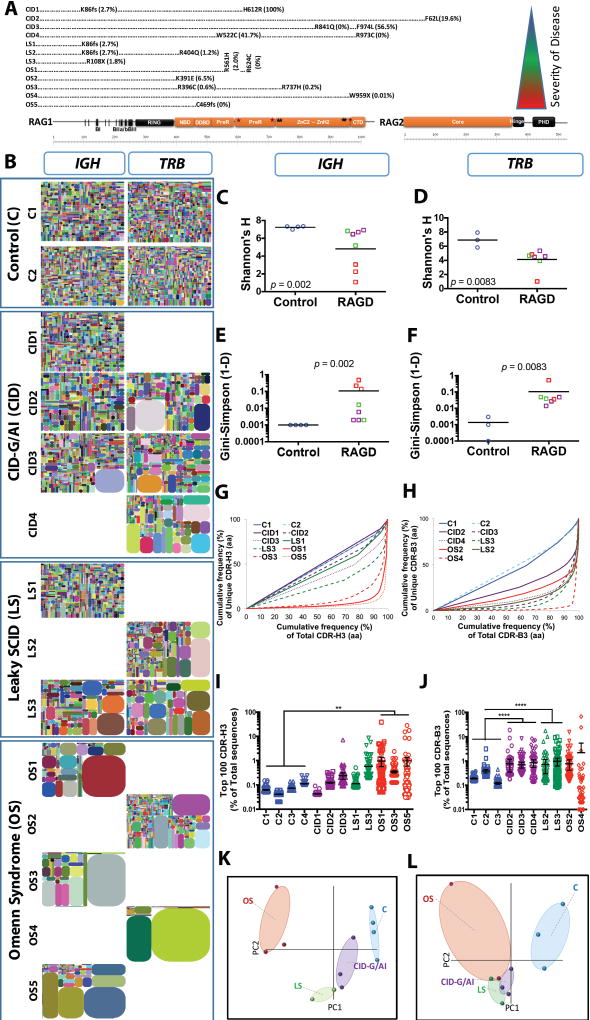
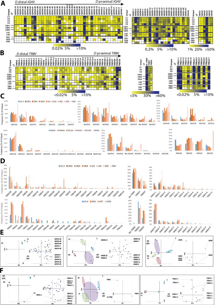
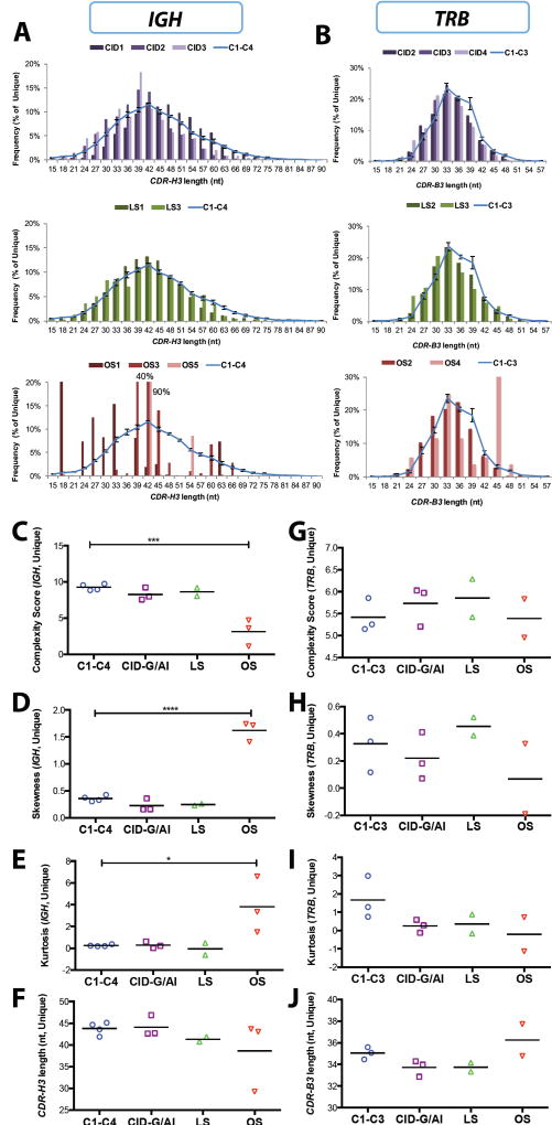
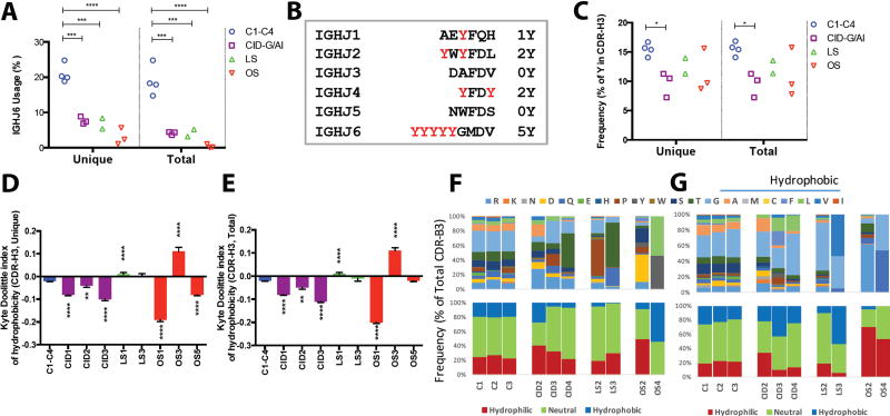
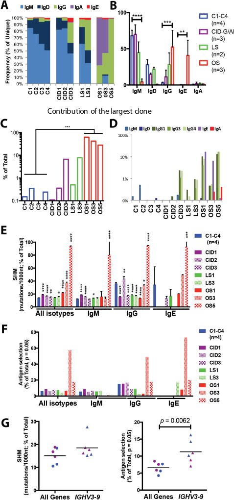
Recombination-activating genes 1 and 2 (RAG1 and RAG2) play a critical role in T and B cell development by initiating the recombination process that controls the expression of T cell receptor (TCR) and immunoglobulin genes. Mutations in the RAG1 and RAG2 genes in humans cause a broad spectrum of phenotypes, including severe combined immunodeficiency (SCID) with lack of T and B cells, Omenn syndrome, leaky SCID, and combined immunodeficiency with granulomas or autoimmunity (CID-G/AI). Using next-generation sequencing, we analyzed the TCR and B cell receptor (BCR) repertoire in 12 patients with RAG mutations presenting with Omenn syndrome (n = 5), leaky SCID (n = 3), or CID-G/AI (n = 4). Restriction of repertoire diversity skewed usage of variable (V), diversity (D), and joining (J) segment genes, and abnormalities of CDR3 length distribution were progressively more prominent in patients with a more severe phenotype. Skewed usage of V, D, and J segment genes was present also within unique sequences, indicating a primary restriction of repertoire. Patients with Omenn syndrome had a high proportion of class-switched immunoglobulin heavy chain transcripts and increased somatic hypermutation rate, suggesting in vivo activation of these B cells. These data provide a framework to better understand the phenotypic heterogeneity of RAG deficiency.


Figures

Fig. 1
Fig. 1. Progressive IGH and TRB repertoire restriction with increased clonality in the patients with RAG deficiency
Schematic representation of RAG1 and RAG2 protein with the mutations of the 12 patients according to the severity of clinical presentation from top to bottom (A). Tree maps representing the diversity and clonality of IGH and TRB (B) repertoires from healthy donor controls (representative data from two subjects are presented) and patients with RAG mutations. Each dot represents a unique V to J joining, and the size of the dot represents the relative frequency of that rearrangement in the entire population. No amplification products were obtained for IGH repertoire from patients CID4, LS2, OS2, and OS4, and for TRB repertoire for patients CD1, LS1, OS1, OS3, and OS5. Quantification of the diversity (C, D) and unevenness (E, F) of the IGH (C, E) and TRB (D, F) repertoires using Shannon’s H index of diversity and Gini- Sipmson’s index of unevenness in healthy controls (blue circles), and patients with CID-G/AI (purple boxes), LS (green boxes), and OS (red boxes). The cumulative frequencies of unique versus total CDR3 clonotypes are shown for IGH (G) and TRB (H) repertoires (CDR-H3 and CDR-B3, respectively). Mean values ± SE are shown; t-test was used for statistical analysis. Representation of the frequency of the top 100 most abundant clones for IGH (I) and TRB (J) sequences in RAG-mutated patients and healthy controls (mean ± SE; ANOVA with post hoc test of Dunnett’s multiple comparisons with *** 0.001 < p < 0.01 and * p < 0.05). Sample plots illustrating the segregation of the various patient groups from healthy controls based on primary component (PC) 1 and 2 determined by five variables (RAG recombination activity, Shannon’s H, Gini-Simpson, number of total and unique sequences) for the IGH (K) and TRB (L) repertoires.
Fig. 2
Fig. 2. Differential usage of V, D and J genes in the IGH and TRB repertoires of patients with RAG deficiency
Heat map representing the frequency of V, D and J gene usage among unique IGH (A) and TRB (B) sequences from healthy controls and RAG-mutated patients. Relative frequency of usage of IGHV and IGHD gene families, and of individual IGHJ genes, in healthy controls and in patients (C, upper panel) and in Abelson virus-transformed pro-B cell lines expressing various RAG1 mutations (C, lower panel). Relative frequency of usage of TRBV gene family and of TRBD and TRBJ genes in healthy controls and in patients (D, upper panel) and in iPS-derived thymocytes (D, lower panel). In panel E, differential usage of IGHV, IGHD and IGHJ genes, segregating control and patient samples and the various genes according to PC1 and PC2 is shown as sample plots (left panels) and variable plots (right panels). In panel F, differential usage of TRBV, TRBD and TRB genes, segregating control and patient samples and the various genes according to PC1 and PC2 is shown as sample plots (left panels) and variable plots (right panels).
Fig. 3
Fig. 3. Characteristics of the CDR3 region of IGH and TRB unique sequences in peripheral blood lymphocytes
Distribution of the length of the CDR3 region of IGH (CDR-H3) (A) and TRB (CDR-B3) (B) unique sequences from peripheral blood of patients with RAG deficiency and healthy controls (C1-C4; C6-C8). In panels A and B, the distribution of the CDR3 length in healthy controls is depicted as a blue line (representing mean values ± SE). Complexity scores (C, G), skewness (D, H), kurtosis (E, I) and average length in nucleotides (nt) (F, J) of the IGH (C-F) and TRB (G-J) CDR3 unique sequences in patients with RAG deficiency and controls. In panels C-J, for each group, mean values are shown, and statistical significance was assessed by ANOVA.
Fig. 4
Fig. 4. Abnormal amino acid composition of CDR3 in the IGH and TRB sequences
Frequency of usage of the IGHJ6 gene among unique CDR-H3 sequences (A). Summary of the Y content in the IGHJ genes (B). Percentage of tyrosine residues in the CDR-H3 of unique and total sequences (C). Summary of CDR-H3 hydrophobicity profile depicted as average Kyte-Doolittle index of hydrophobicity (mean ± SE) in patients and healthy control blood samples for unique (D) and total (E) sequences. (**, p<0.01; ***, p <0.001; ****, p < 0.0001; one-tail unpaired t-test). Amino acid composition of CDR-B3 in patients and healthy control for amino acid positions 6 (F) and 7 (G) of the 13 aa-long CDR-B3.
Fig. 5
Fig. 5. Distribution of immunoglobulin heavy chain isotypes, somatic hypermutation and antigen-driven selection in peripheral blood B cells of patients with RAG deficiency
Frequency of immunoglobulin heavy chain constant gene usage among unique IGH sequences from peripheral blood lymphocytes of RAG-deficient patients and healthy controls (A and B). In panel B, mean values ± SE are shown (one-tail, unpaired t-test). Contribution of the most abundant clonotype to the total number of IGH sequences in patients and controls (C). Distribution of various isotypes among the most abundant IGH transcript (D). Rate of somatic hypermutation (SHM) in IGH transcripts (E, mean ± SE; unpaired t-test). Frequency of unique IGH transcripts displaying evidence of antigen-mediated selection based on the distribution of replacement and silent mutations (F). Rate of SHM and antigen mediated selection in IGHV3-9 for patients with CID-G/AI and healthy controls with line at the mean (G).
Fig. 6
Fig. 6. Mapping the disease-related mutations onto the synaptic-RAG complex models
Overview of the disease-related mutations shown as space filling models mapped onto the ribbon diagram of the synaptic RAG complex structure (PDB ID 3JBY, top and bottom view) (A). Residues in zebrafish rag1 and rag2 and the equivalent residues that have been mutated in patients are labeled. Only one RAG1-RAG2 subunit is labeled for explicitness on the side view. Labeled in purple and red are residues that are mutated in patients with CID-G/AI and OS, respectively. Residues affected by mutations that correspond to the allele with lower recombination activity in compound heterozygous patients are labeled in black. Examples of the detailed interactions between the equivalent residues from patients and the RSS intermediates or partner residues (PBD ID 3GNA and 3JBY) (B). Equivalent residues that have interaction with RSS intermediates are shown as sticks and highlighted in magenta. The nucleotides in the RSS intermediates that have interaction with protein residues are shown as sticks and highlighted in cyan. The partner residues are shown as sticks and highlighted in marine. Potential interactions are displayed as red dashed lines. z is abbreviated for zebrafish and m is for mouse. All molecular representations were generated in PyMOL (http://www.pymol.org) (47).

References

    1. Villa A, Santagata S, Bozzi F, Giliani S, Frattini A, Imberti L, Gatta LB, Ochs HD, Schwarz K, Notarangelo LD, Vezzoni P, Spanopoulou E. Partial V(D)J recombination activity leads to Omenn syndrome. Cell. 1998;93:885–896. - PubMed
    1. Shearer WT, Dunn E, Notarangelo LD, Dvorak CC, Puck JM, Logan BR, Griffith LM, Kohn DB, O'Reilly RJ, Fleisher TA, Pai SY, Martinez CA, Buckley RH, Cowan MJ. Establishing diagnostic criteria for severe combined immunodeficiency disease (SCID), leaky SCID, and Omenn syndrome: the Primary Immune Deficiency Treatment Consortium experience. J Allergy Clin Immunol. 2014;133:1092–1098. - PMC - PubMed
    1. de Villartay JP, Lim A, Al-Mousa H, Dupont S, Dechanet-Merville J, Coumau-Gatbois E, Gougeon ML, Lemainque A, Eidenschenk C, Jouanguy E, Abel L, Casanova JL, Fischer A, Le Deist F. A novel immunodeficiency associated with hypomorphic RAG1 mutations and CMV infection. J Clin Invest. 2005;115:3291–3299. - PMC - PubMed
    1. Ehl S, Schwarz K, Enders A, Duffner U, Pannicke U, Kuhr J, Mascart F, Schmitt-Graeff A, Niemeyer C, Fisch P. A variant of SCID with specific immune responses and predominance of gamma delta T cells. J Clin Invest. 2005;115:3140–3148. - PMC - PubMed
    1. Schuetz C, Huck K, Gudowius S, Megahed M, Feyen O, Hubner B, Schneider DT, Manfras B, Pannicke U, Willemze R, Knuchel R, Gobel U, Schulz A, Borkhardt A, Friedrich W, Schwarz K, Niehues T. An immunodeficiency disease with RAG mutations and granulomas. N Engl J Med. 2008;358:2030–2038. - PubMed