. 2017 Aug 10;548(7666):239-243.
doi: 10.1038/nature23297. Epub 2017 Aug 2.

A Braf kinase-inactive mutant induces lung adenocarcinoma

Affiliations

A Braf kinase-inactive mutant induces lung adenocarcinoma

Patricia Nieto et al. Nature. .

Abstract

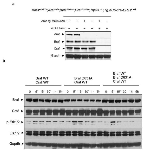
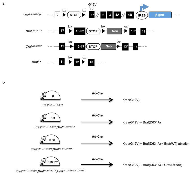
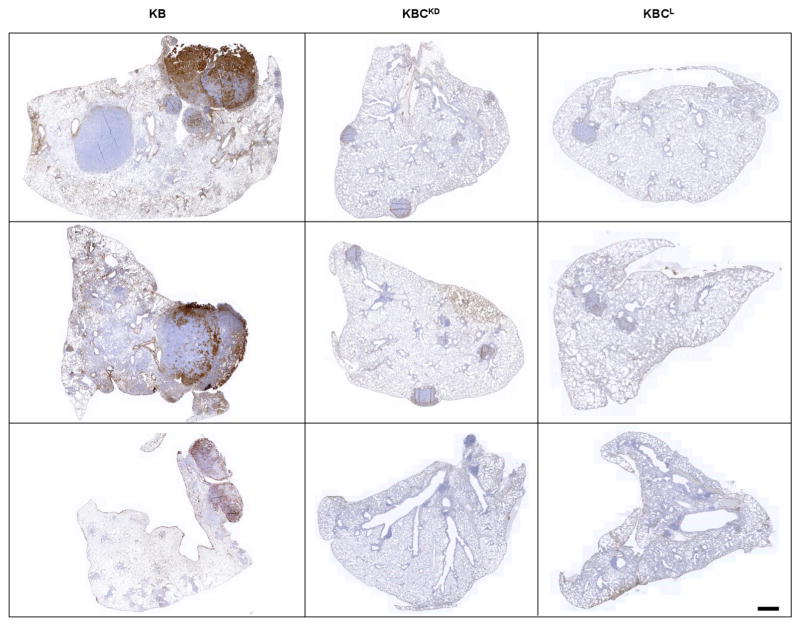
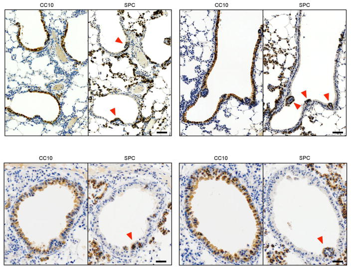
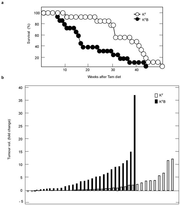
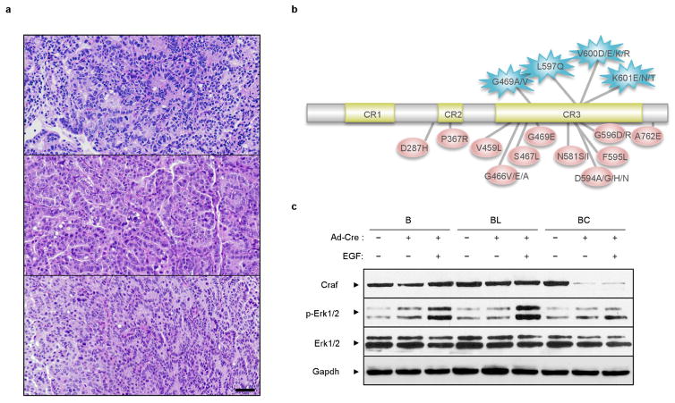
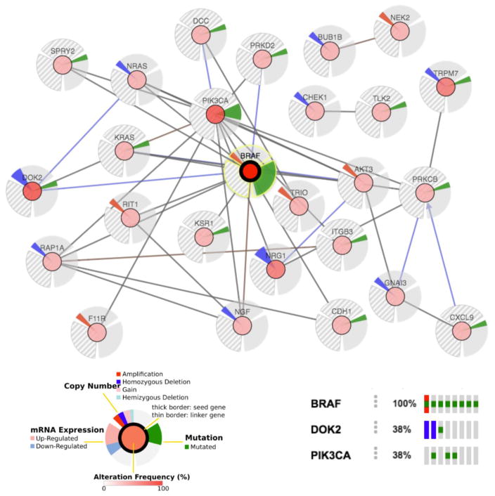
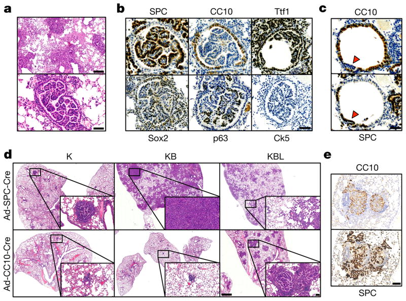
The initiating oncogenic event in almost half of human lung adenocarcinomas is still unknown, a fact that complicates the development of selective targeted therapies. Yet these tumours harbour a number of alterations without obvious oncogenic function including BRAF-inactivating mutations. Inactivating BRAF mutants in lung predominate over the activating V600E mutant that is frequently observed in other tumour types. Here we demonstrate that the expression of an endogenous Braf(D631A) kinase-inactive isoform in mice (corresponding to the human BRAF(D594A) mutation) triggers lung adenocarcinoma in vivo, indicating that BRAF-inactivating mutations are initiating events in lung oncogenesis. Moreover, inactivating BRAF mutations have also been identified in a subset of KRAS-driven human lung tumours. Co-expression of Kras(G12V) and Braf(D631A) in mouse lung cells markedly enhances tumour initiation, a phenomenon mediated by Craf kinase activity, and effectively accelerates tumour progression when activated in advanced lung adenocarcinomas. We also report a key role for the wild-type Braf kinase in sustaining Kras(G12V)/Braf(D631A)-driven tumours. Ablation of the wild-type Braf allele prevents the development of lung adenocarcinoma by inducing a further increase in MAPK signalling that results in oncogenic toxicity; this effect can be abolished by pharmacological inhibition of Mek to restore tumour growth. However, the loss of wild-type Braf also induces transdifferentiation of club cells, which leads to the rapid development of lethal intrabronchiolar lesions. These observations indicate that the signal intensity of the MAPK pathway is a critical determinant not only in tumour development, but also in dictating the nature of the cancer-initiating cell and ultimately the resulting tumour phenotype.


Conflict of interest statement

The authors declare competing financial interests: details are available in the online version of the paper.

Figures

Extended Data Figure 1
Extended Data Figure 1. Braf(D631A)-dependent activation of MAPK signalling in vitro
a, Western blot analysis of the expression levels of the three Raf isoforms in lysates derived from Kras+/G12V;Araf+/+;Braf lox/lox; Craf lox/lox;Trp53−/−;Tg.hUb-cre-ERT2+/T lung adenocarcinoma cell lines. The endogenous Araf alleles were eliminated using CRISPR–Cas9 editing where indicated. Braf lox and Craf lox alleles were eliminated by addition of 4-hydroxytamoxifen (4-OHT) to induce their Cre-mediated recombination where indicated. Gapdh is shown as a loading control. b, Kras+/G12V;A-Raf−/−;Braf lox/lox;Craf lox/lox;Trp53−/−;Tg.hUb-cre-ERT2+/T lung adenocarcinoma cell lines were infected with lentiviral particles expressing combinations of wild-type Braf, kinase-inactive Braf(D631A) and wild-type Craf as indicated before the addition of 4-hydroxytamoxifen to induce elimination of the endogenous conditional Braf and Craf alleles. Cells were serum-starved for 24 h and re-stimulated with serum for the indicated times. The activation of MAPK signalling was assessed by western blot analysis of p-Erk1/2. Expression levels of the exogenous Braf and Craf alleles as well as endogenous Erk1/2 and Gapdh are shown as loading controls. For gel source data, see Supplementary Fig. 1.
Extended Data Figure 2
Extended Data Figure 2. Mouse alleles and strains used in the study
a, Expression of the endogenous KrasG12V oncogene is achieved by Cre-mediated excision of the transcriptional STOP cassette preceding the KrasLSLG12Vgeo allele. Cells expressing the oncogene can be identified by X-gal staining owing to the co-expression of a bicistronic β-galactosidase (β-geo) reporter. Likewise, Cre-mediated excision of the lox-STOP-lox cassette (containing the indicated wild-type exons) induces the expression of the kinase-dead Braf(D631A) and Craf(D468A) mutants instead of the wild-type isoforms. Finally, Cre-mediated excision of the conditional Braf lox allele results in the elimination of the wild-type Braf protein. b, Schematic representation of the proteins expressed in lung cells following intratracheal infection with Ad-Cre virus in the compound strains used in the study.
Extended Data Figure 3
Extended Data Figure 3. Activation of p-Erk1/2 in lung adenocarcinomas driven by concomitant expression of Kras(G12V) and Braf(D631A) depends on Craf kinase activity
Representative images of p-Erk1/2 immunostaining of paraffin-embedded lung sections (n =3 per genotype) from Ad-Cre-infected Kras+/LSLG12Vgeo;Braf +/LSLD631A (KB), Kras+/LSLG12Vgeo;Braf +/LSLD631A;Craf LSLD468A/LSLD468A (KBCKD) and Kras+/LSLG12Vgeo;Braf +/LSLD631A;Craf lox/lox (KBCL). Tumour samples were collected 6 months after Ad-Cre intratracheal infection coincident with the humane end point of the KB strain. Scale bar, 1 mm.
Extended Data Figure 4
Extended Data Figure 4. MAPK hyperactivation induces transdifferentiation of the bronchiolar epithelium leading to papillary carcinoma
Immunostaining of paraffin-embedded sections showing transdifferentiation of the bronchiolar epithelium at early stages (1 week) after Ad-Cre infection of Kras+/LSLG12Vgeo;Braf lox/LSLD631A (KBL). Images display staining using antibodies against CC10 (club cell marker, left panels) and SPC (AT2 marker, right panels). Protruding bronchiolar papillary growth is invariably associated with a transdifferentiation process illustrated by the acquisition of SPC+ staining (red arrowheads). Scale bar, 100 μm (top panels), 50 μm (bottom panels).
Extended Data Figure 5
Extended Data Figure 5. Expression of an endogenous Braf(D631A) inactive mutant allele in mice harbouring KrasG12V-driven lung adenocarcinomas detectable by CT increases tumour growth and reduces survival
a, Survival of Kras+/FSFG12V;Braf +/LSLD631A;Tg.hUb-cre-ERT2+/T (KFB, solid circles n = 14) and control Kras+/FSFG12V;Tg.hUb-cre-ERT2+/T (KF, empty circles n = 13) mice bearing lung adenocarcinomas detectable by CT. Upon tumour detection mice were maintained on a tamoxifen-containing diet to activate expression of the Braf(D631A) kinase-dead isoform. P < 0.0058, obtained using the log-rank test (Mantel–Cox). b, Waterfall plot representation of the tumour volume increase (measured as fold change) when re-evaluated by CT after 8 weeks of continuous tamoxifen-containing diet in Kras+/FSFG12V;Braf +/LSLD631A;Tg.hUb-cre-ERT2+/T (KFB, n = 29 tumours from 14 mice) and control Kras+/FSFG12V; Tg.hUb-cre-ERT2+/T (KF, n = 38 tumours from 15 mice) cohorts. The same dataset is represented in Fig. 4a.
Extended Data Figure 6
Extended Data Figure 6. Histology of Braf(D631A)-driven lung adenocarcinoma and evaluation of Braf(D631A)-dependent activation of MAPK signalling in vitro.
a, Haematoxylin and eosin staining of paraffin-embedded lung sections from Ad-Cre-infected Braf +/LSLD631A mice killed at humane end point. Sections display moderately to poorly circumscribed tumours occupying nearly an entire lung lobe, with compressed adjacent lung parenchyma and peripherally infiltrated by mononuclear inflammatory cells, mainly macrophages. Tumour masses are composed by papillary outgrowths or less-differentiated areas with increased cellular pleomorphism. Images correspond to three independent mice. Scale bar, 100 μm. b, Schematic representation of BRAF mutations detected in human tumours with high (top) or low/absent (bottom) kinase activity when compared to the wild-type isoform (see also the accompanying manuscript by Yao et al.). c, Primary keratinocytes derived from Braf +/LSLD631A (B), Braf lox/LSLD631A (BL) and Braf +/LSLD631A; Craf lox/lox (BC) mice were infected with Ad-Cre (5 MOI) and treated with recombinant EGF (150 ng ml−1 for 1 h) where indicated. The activation of MAPK signalling was assessed by western blot analysis of p-Erk1/2, demonstrating that the activation of MAPK signalling by Braf(D631A) in epithelial cells requires upstream RTK activation and is enhanced by elimination of the wild-type Braf allele. Efficient recombination of the Craf conditional alleles in Ad-Cre-infected BC cells is also shown, demonstrating that MAPK activation by Braf(D631A) is Craf-dependent. Expression levels of Erk1/2 and Gapdh are shown as loading controls. For gel source data, see Supplementary Fig. 1.
Extended Data Figure 7
Extended Data Figure 7. Co-occurring alterations in lung adenocarcinoma patients with BRAF-inactivating mutations
Whole-genome sequencing data available from the TCGA LUAD study at cBioportal (http://www.cbioportal.org) was used to generate a gene network for the analysis of genetic alterations coincident with BRAF hypoactive mutations. PIK3CA mutations (Q296E, E542K and E545K) were the most frequent co-occurring event. In addition, mutations or gene deletions in known RTK signalling antagonists (DOK2, SPRY2), MAPK scaffolds (KSR1), members of the RAS subfamily (RAP1A, RIT1) or the guanine nucleotide exchange factor TRIO might potentially cooperate with inactive BRAF to sustain MAPK activity.
Figure 1
Figure 1. Combinations of KrasG12V, Braf D631A and wild-type Braf alleles establish a MAPK activity window that determines cell transformation and oncogene toxicity
a, Whole-mount X-gal staining of representative lung sections (n = 5 per genotype) from Kras+/LSLG12Vgeo (K), Kras+/LSLG12VgeoBraf ; +/LSLD631A (KB) and Kras+/LSLG12VgeoBraf ; lox/LSLD631A (KBL) mice 1 month after Ad-Cre infection. X-gal staining identifies β-galactosidase expression as a surrogate marker for KrasG12V-expressing cells. Scale bar, 1mm. Insets show high-magnification images. Scale bar, 100 μm. b, Western blot analysis of lung lysates from Kras+/LSLG12Vgeo (K), Kras+/LSLG12Vgeo;Braf +/LSLD631A (KB) and Kras+/LSLG12Vgeo;Braf lox/LSLD631A (KBL) mice 1 week after Ad-Cre infection. Migration of p19ARF, p53, γ-H2AX, cleaved caspase-3 (C3A), p-Erk1/2, Erk1/2, p-p90Rsk and p90Rsk is indicated by arrowheads. Gapdh was used as loading control. Lysates from two independent animals per genotype are shown. c, Representative immunostaining of paraffin-embedded lung sections (n = 5 per genotype) from Kras+/LSLG12Vgeo (K), Kras+/LSLG12Vgeo;Braf +/LSLD631A (KB) and Kras+/LSLG12Vgeo;Braf lox/LSLD631A (KBL) mice 1 week after Ad-Cre infection using the indicated antibodies. Scale bar, 50 μm. d, Whole- mount X-gal staining of representative lung sections (n = 3 per genotype) from Kras+/LSLG12Vgeo (K), Kras+/LSLG12Vgeo;Braf +/LSLD631A (KB) and Kras+/LSLG12VgeoBraf ; lox/LSLD631A (KBL) mice 1 month after infection with 108 Ad-Cre particles. During this period, mice were treated with the indicated doses of the Mek inhibitor PD-0325901. The percentage of KrasG12V-expressing cells (X-gal-positive area) per lung section is indicated in each panel (top left, shown as mean ± s.d.). Scale bar, 200 μm. Insets in each panel show high-magnification images (top right; scale bar, 50 μm) or representative images of p-Erk1/2 immunostaining (bottom right; scale bar, 25 μm).
Figure 2
Figure 2. Mice concomitantly expressing Kras(G12V) and Braf(D631A) show reduced survival and increased tumour development mediated by Craf kinase activity
a, Survival of Kras+/LSLG12Vgeo (K, open circles, n =14), Kras +/LSLG12Vgeo;Braf +/LSLD631A (KB, solid circles, n =22) and Kras+/LSLG12Vgeo;Braf lox/LSLD631A (KBL, open triangles, n = 17) mice after Ad-Cre intratracheal infection. P < 0.0001, obtained using the log-rank test (Mantel–Cox). b, Quantification of average tumour (adenoma and adenocarcinoma) burden (per cent of total lung area) in lung sections from Kras+/LSLG12Vgeo (K, n =5), Kras+/LSLG12Vgeo;Braf +/LSLD631A (KB, n =7) and Kras+/LSLG12Vgeo;Braf lox/LSLD631A (KBL, n = 7) mice 6 months after Ad-Cre intratracheal infection. **P <0.01, *P <0.05. c, Representative haematoxylin and eosin staining of paraffin-embedded lung sections obtained from Kras+/LSLG12Vgeo (K, n =5), Kras+/LSLG12Vgeo;Braf +/LSLD631A (KB, n =7) and Kras+/LSLG12Vgeo;Braf lox/LSLD631A (KBL, n = 7) mice 6 months after Ad-Cre infection. Scale bar, 5 mm. d, Representative immunostaining of paraffin-embedded sections showing tumours from Ad-Cre-infected Kras+/LSLG12Vgeo;Braf +/LSLD631A (KB, n = 7) mice killed at humane end point using antibodies against SPC and CC10. Scale bar, 1 mm. e, Quantification of average tumour (adenoma and adenocarcinoma) burden (per cent of total lung area) in lung sections from Kras+/LSLG12Vgeo;Braf +/LSLD631A (KB, n = 7) and Kras+/LSLG12VgeoBraf ; +/LSLD631ACraf ; LSLD468A/LSLD468A (KBCKD, n = 5) mice 6 months after Ad-Cre intratracheal infection. **P <0.01. f, Representative haematoxylin and eosin staining of paraffin-embedded lung sections (n = 5 per genotype) from Kras+/LSLG12Vgeo;Braf +/LSLD631A (KB), and Kras+/LSLG12Vgeo;Braf +/LSLD631A;Craf LSLD468A/LSLD468A (KBCKD) mice 6 months after Ad-Cre infection. Scale bar, 5 mm. g, Survival of Kras+/LSLG12Vgeo;Braf +/LSLD631A (KB, solid circles, n =22), Kras+/LSLG12Vgeo;Braf +/LSLD631ACraf ; LSLD468A/LSLD468A (KBCKD, solid triangles, n = 12) and Kras+/LSLG12Vgeo;Braf +/LSLD631A;Craf lox/lox (KBCL, empty squares, n = 7) mice after Ad-Cre intratracheal infection. P <0.0001, obtained using the log-rank test (Mantel–Cox).
Figure 3
Figure 3. Hyperactive MAPK signalling triggers bronchiolar carcinoma by transdifferentiation of club cells
a, Haematoxylin and eosin staining of paraffin-embedded sections showing the presence of intrabronchiolar carcinoma in Ad-Cre-infected Kras+/LSLG12Vgeo;Braf lox/LSLD631A (KBL) mice killed at humane end point. Two different magnifications are shown. Scale bar, 200 μm (top) and 50 μm (bottom). b, Representative immunostaining of paraffin-embedded sections showing intrabronchiolar tumours from Ad-Cre-infected Kras+/LSLG12Vgeo;Braf lox/LSLD631A (KBL, n = 5) mice using antibodies against, SPC, CC10, Ttf1, Sox2, p63 and Ck5. Scale bar, 50 μm. c, Immunostaining of paraffin-embedded consecutive sections showing early intrabronchiolar lesions (1 week after Ad-Cre infection) in Kras+/LSLG12Vgeo;Braf lox/LSLD631A (KBL) mice using antibodies against SPC and CC10. Arrowheads indicate protruding bronchiolar papillary growth undergoing transdifferentiation. Scale bar, 50 μm. d, Representative haematoxylin and eosin staining of paraffin-embedded lung sections (n = 3 per genotype) from Kras+/LSLG12Vgeo (K), Kras+/LSLG12Vgeo; Braf +/LSLD631A (KB) and Kras+/LSLG12Vgeo;Braf lox/LSLD631A (KBL) mice killed 2 months after infection with cell-type-restricted Ad-Cre viruses that selectively induce Cre-mediated recombination in AT2 (Ad-SPC-Cre) (top) and club cells (Ad-CC10-Cre) (bottom). Scale bar, 500 μm. Insets show high-magnification images. Scale bar, 100 μm. e, SPC and CC10 immunostaining of paraffin-embedded consecutive sections showing intrabronchiolar tumours from Kras+/LSLG12Vgeo;Braf lox/LSLD631A (KBL) mice infected with lineage-specific Ad-CC10-Cre. Scale bar, 100 μm.
Figure 4
Figure 4. Activation of endogenous Braf D631A results in lung adenocarcinoma development
a , Kras+/FSFG12V;Braf +/LSLD631A; Tg.hUb-cre-ERT2+/T (KFB) and control Kras+/FSFG12V;Tg.hUb-cre-ERT2+/T (KF) mice were infected intratracheally with Ad-Flp and tumour formation was monitored by computed tomography (CT). Mice bearing tumours visible on CT (KFB, n = 29 tumours from 14 mice and KF, n =38 tumours from 15 mice) were fed ad libitum a tamoxifen-containing diet resulting in expression of the Braf(D631A) kinase-dead isoform in KFB. Tumour volume increase (measured as fold change) was re-evaluated by CT after 8 weeks in continuous diet. **P <0.01. b, Haematoxylin and eosin (H&E) staining, as well as SPC and CC10 immunostaining, of paraffin-embedded lung sections from Ad-Cre-infected Braf +/LSLD631A mice killed at humane end-point. Scale bar, 500 μm and 50 μm (top right). c, Representative images of p-Erk1/2 immunostaining of paraffin-embedded lung sections (n = 3 per genotype) from Ad-Cre-infected Kras+/LSLG12Vgeo, Kras+/LSLG12Vgeo;Trp53 lox/lox, Braf +/LSLV637E (equivalent to human BRAFV600E) or Braf +/LSLD631A mice. Scale bar, 100 μm.

Comment in

References

    1. Chang MT, et al. Identifying recurrent mutations in cancer reveals widespread lineage diversity and mutational specificity. Nat Biotechnol. 2016;34:155–163. - PMC - PubMed
    1. Heidorn SJ, et al. Kinase-dead BRAF and oncogenic RAS cooperate to drive tumor progression through CRAF. Cell. 2010;140:209–221. - PMC - PubMed
    1. Poulikakos PI, Zhang C, Bollag G, Shokat KM, Rosen N. RAF inhibitors transactivate RAF dimers and ERK signalling in cells with wild-type BRAF. Nature. 2010;464:427–430. - PMC - PubMed
    1. Sebolt-Leopold JS, Herrera R. Targeting the mitogen-activated protein kinase cascade to treat cancer. Nat Rev Cancer. 2004;4:937–947. - PubMed
    1. Rauen KA. The RASopathies. Annu Rev Genomics Hum Genet. 2013;14:355–369. - PMC - PubMed

Publication types