

PKC-epsilon deficiency alters progenitor cell populations in favor of megakaryopoiesis
- PMID: 28783756
- PMCID: PMC5544228
- DOI: 10.1371/journal.pone.0182867
PKC-epsilon deficiency alters progenitor cell populations in favor of megakaryopoiesis
Abstract
Background: It has long been postulated that Protein Kinase C (PKC) is an important regulator of megakaryopoiesis. Recent contributions to the literature have outlined the functions of several individual PKC isoforms with regard to megakaryocyte differentiation and platelet production. However, the exact role of PKCε remains elusive.
Objective: To delineate the role of PKCε in megakaryopoiesis.
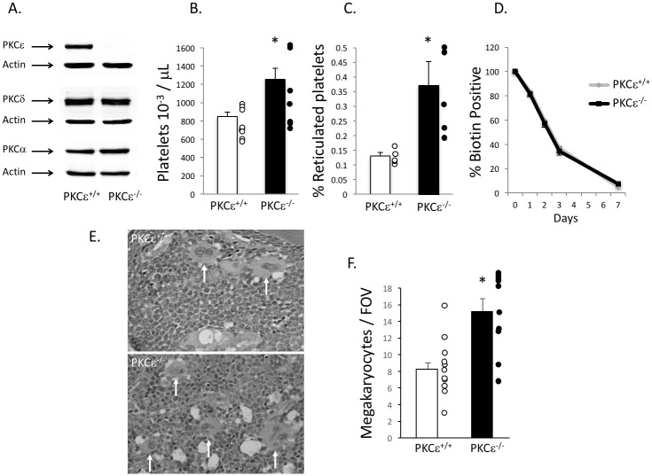
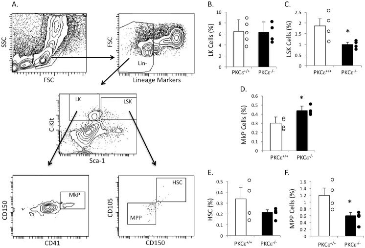
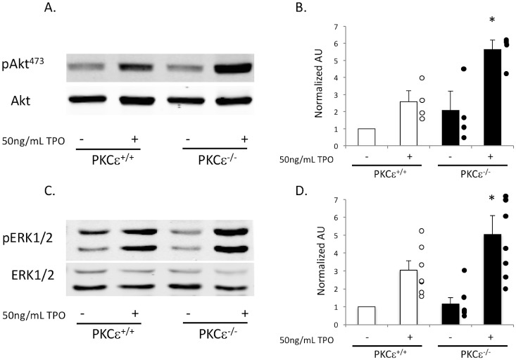
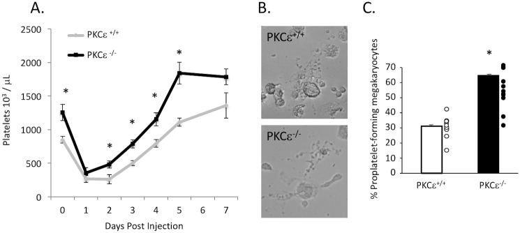
Approach and results: We used a PKCε knockout mouse model to examine the effect of PKCε deficiency on platelet mass, megakaryocyte mass, and bone marrow progenitor cell distribution. We also investigated platelet recovery in PKCε null mice and TPO-mediated signaling in PKCε null megakaryocytes. PKCε null mice have higher platelet counts due to increased platelet production compared to WT littermate controls (p<0.05, n = 8). Furthermore, PKCε null mice have more bone marrow megakaryocyte progenitor cells than WT littermate control mice. Additionally, thrombopoietin-mediated signaling is perturbed in PKCε null mice as Akt and ERK1/2 phosphorylation are enhanced in PKCε null megakaryocytes stimulated with thrombopoietin. Finally, in response to immune-induced thrombocytopenia, PKCε null mice recovered faster and had higher rebound thrombocytosis than WT littermate control mice.
Conclusions: Enhanced platelet recovery could be due to an increase in megakaryocyte progenitor cells found in PKCε null mice as well as enhanced thrombopoietin-mediated signaling observed in PKCε deficient megakaryocytes. These data suggest that PKCε is a negative regulator of megakaryopoiesis.
Conflict of interest statement
Figures
Similar articles
-
Protein kinase C δ deficiency enhances megakaryopoiesis and recovery from thrombocytopenia.Arterioscler Thromb Vasc Biol. 2014 Dec;34(12):2579-85. doi: 10.1161/ATVBAHA.114.304492. Epub 2014 Oct 30. Arterioscler Thromb Vasc Biol. 2014. PMID: 25359855 Free PMC article.
-
Roles of focal adhesion kinase (FAK) in megakaryopoiesis and platelet function: studies using a megakaryocyte lineage specific FAK knockout.Blood. 2008 Jan 15;111(2):596-604. doi: 10.1182/blood-2007-05-089680. Epub 2007 Oct 9. Blood. 2008. PMID: 17925492 Free PMC article.
-
Humanized VB22B minibody for human Mpl stimulates human megakaryopoiesis but does not enhance platelet aggregation.Exp Hematol. 2011 Aug;39(8):829-36. doi: 10.1016/j.exphem.2011.05.001. Epub 2011 May 7. Exp Hematol. 2011. PMID: 21605620
-
Physiologic role of TPO in thrombopoiesis.Stem Cells. 1996;14 Suppl 1:133-8. doi: 10.1002/stem.5530140717. Stem Cells. 1996. PMID: 11012213 Review.
-
Milestones in understanding platelet production: a historical overview.Br J Haematol. 2014 Apr;165(2):248-58. doi: 10.1111/bjh.12781. Epub 2014 Feb 14. Br J Haematol. 2014. PMID: 24528208 Review.
Cited by
-
micro-RNAs dependent regulation of DNMT and HIF1α gene expression in thrombotic disorders.Sci Rep. 2019 Mar 20;9(1):4815. doi: 10.1038/s41598-018-38057-6. Sci Rep. 2019. PMID: 30894555 Free PMC article.
-
Unraveling the impact of crizotinib to promote megakaryopoiesis for alleviating thrombocytopenia in myelodysplastic neoplasms.Leukemia. 2025 Aug 14. doi: 10.1038/s41375-025-02729-w. Online ahead of print. Leukemia. 2025. PMID: 40813622
-
PKCδ reveals a tumor promoter function by promoting cell proliferation and migration in somatotropinomas.Int J Clin Exp Pathol. 2018 Jan 1;11(1):208-215. eCollection 2018. Int J Clin Exp Pathol. 2018. PMID: 31938102 Free PMC article.
-
Nootkatone Derivative Nootkatone-(E)-2-iodobenzoyl hydrazone Promotes Megakaryocytic Differentiation in Erythroleukemia by Targeting JAK2 and Enhancing JAK2/STAT3 and PKCδ/MAPK Crosstalk.Cells. 2024 Dec 26;14(1):10. doi: 10.3390/cells14010010. Cells. 2024. PMID: 39791711 Free PMC article.
-
Electroacupuncture Regulates Pain Transition Through Inhibiting PKCε and TRPV1 Expression in Dorsal Root Ganglion.Front Neurosci. 2021 Jul 20;15:685715. doi: 10.3389/fnins.2021.685715. eCollection 2021. Front Neurosci. 2021. PMID: 34354561 Free PMC article.
References
-
- Travlos GS. Normal structure, function, and histology of the bone marrow. Toxicol Pathol. 2006;34(5):548–65. doi: 10.1080/01926230600939856 - DOI - PubMed
-
- Broudy VC, Lin NL, Kaushansky K. Thrombopoietin (c-mpl ligand) acts synergistically with erythropoietin, stem cell factor, and interleukin-11 to enhance murine megakaryocyte colony growth and increases megakaryocyte ploidy in vitro. Blood. 1995;85(7):1719–26. Epub 1995/04/01. - PubMed
-
- Teramura M, Kobayashi S, Hoshino S, Oshimi K, Mizoguchi H. Interleukin-11 enhances human megakaryocytopoiesis in vitro. Blood. 1992;79(2):327–31. Epub 1992/01/15. - PubMed
-
- Drachman JG, Millett KM, Kaushansky K. Thrombopoietin signal transduction requires functional JAK2, not TYK2. J Biol Chem. 1999;274(19):13480–4. Epub 1999/05/01. - PubMed
MeSH terms
Substances
Grants and funding
LinkOut - more resources
Full Text Sources
Other Literature Sources
Medical
Molecular Biology Databases
Miscellaneous
