

A superficial esophageal cancer in an epiphrenic diverticulum treated by endoscopic submucosal dissection
- PMID: 28784105
- PMCID: PMC5547519
- DOI: 10.1186/s12876-017-0649-y
A superficial esophageal cancer in an epiphrenic diverticulum treated by endoscopic submucosal dissection
Abstract
Background: We report a unique case of a superficial esophageal cancer arising in a single diverticulum, diagnosed with magnifying image-enhanced endoscopy and then successfully treated by endoscopic submucosal dissection (ESD).
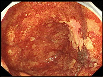
Case presentation: A 66-year-old man with alcohol-related liver injury visited our hospital for endoscopy for investigation of varix. Esophagogastroduodenoscopy showed no varix but a large epiphrenic diverticulum with an area of fainted redness just above the esophagogastric junction. Narrow band imaging revealed a sharply demarcated brownish dotted area, and dilated intra-epithelial papillary capillary loops (IPCL) were subsequently seen after magnification. Chromoendoscopy with 1% Lugol's iodine solution demonstrated a well-demarcated unstained area, approximately 20 mm in diameter. Endoscopic biopsy revealed a squamous cell carcinoma (SCC).
Conclusion: The tumor was completely resected by ESD without perforation. Histologically, it was an intraepithelial SCC without lympho-vascular invasion of cancer cells. No local recurrence or metastasis was detected at the last follow-up of 42 months.
Keywords: Endoscopic submucosal dissection; Epiphrenic diverticulum; Magnifying endoscopy; Narrow band imaging; Superficial esophageal cancer.
Conflict of interest statement
Ethics approval and consent to participate
Written consent was obtained from the patient. As a case report, approval from the institutional review board was not needed.
Consent for publication
Written informed consent was obtained from the patient for publication of this case report.
Competing interests
The authors declare that they have no competing interests.
Publisher’s Note
Springer Nature remains neutral with regard to jurisdictional claims in published maps and institutional affiliations.
Figures


Similar articles
-
Endoscopic submucosal dissection for superficial esophageal cancer.Dis Esophagus. 2018 Jul 1;31(7). doi: 10.1093/dote/doy021. Dis Esophagus. 2018. PMID: 29982386 Review.
-
Traction-assisted esophageal endoscopic submucosal dissection for treatment of squamous cell carcinoma involving a diverticulum.Dig Endosc. 2019 Jan;31(1):e7-e8. doi: 10.1111/den.13273. Epub 2018 Oct 14. Dig Endosc. 2019. PMID: 30187961 No abstract available.
-
Esophageal diverticulum exposed during endoscopic submucosal dissection of superficial cancer.World J Gastroenterol. 2015 Mar 14;21(10):3121-6. doi: 10.3748/wjg.v21.i10.3121. World J Gastroenterol. 2015. PMID: 25780314 Free PMC article.
-
Endoscopic mucosal resection of early esophageal carcinoma--experience of 9 cases.J Chin Med Assoc. 2008 Jul;71(7):347-52. doi: 10.1016/S1726-4901(08)70137-0. J Chin Med Assoc. 2008. PMID: 18653397
-
Early esophageal squamous cell carcinoma management through endoscopic submucosal dissection.Rev Gastroenterol Mex (Engl Ed). 2018 Jul-Sep;83(3):259-267. doi: 10.1016/j.rgmx.2017.12.004. Epub 2018 Mar 16. Rev Gastroenterol Mex (Engl Ed). 2018. PMID: 29551245 Review. English, Spanish.
Cited by
-
Atypical presentation of an epiphrenic esophageal diverticulum 20 years post fundoplication: a case report and review.J Surg Case Rep. 2024 May 29;2024(5):rjae316. doi: 10.1093/jscr/rjae316. eCollection 2024 May. J Surg Case Rep. 2024. PMID: 38872729 Free PMC article.
-
Early oesophageal carcinoma with a defect of the oesophageal muscularis propria: a rare case report.J Int Med Res. 2023 Oct;51(10):3000605231204422. doi: 10.1177/03000605231204422. J Int Med Res. 2023. PMID: 37903318 Free PMC article.
-
Early esophageal squamous cell carcinoma and diverticulum were cured simultaneously by endoscopic submucosal dissection in a patient.Endoscopy. 2023 Dec;55(S 01):E468-E469. doi: 10.1055/a-2020-9828. Epub 2023 Feb 24. Endoscopy. 2023. PMID: 36828023 Free PMC article. No abstract available.
-
A thoracoscopically resected case of the diverticulum in the middle esophagus.Surg Case Rep. 2019 Jul 9;5(1):109. doi: 10.1186/s40792-019-0668-8. Surg Case Rep. 2019. PMID: 31289952 Free PMC article.
References
-
- Muto M, Minashi K, Yano T, Saito Y, Oda I, Nonaka S, et al. Early detection of superficial squamous cell carcinoma in the head and neck region and esophagus by narrow band imaging: a multicenter randomized controlled trial. J Clin Oncol. 2010;28:1566–1572. doi: 10.1200/JCO.2009.25.4680. - DOI - PMC - PubMed
-
- Oyama T, Inoue H, Arima M, Momma K, Omori T, Ishihara R, et al. Prediction of the invasion depth of superficial squamous cell carcinoma based on microvessel morphology: magnifying endoscopic classification of the Japan esophageal society. Esophagus. 2017;14:105–112. doi: 10.1007/s10388-016-0527-7. - DOI - PMC - PubMed
-
- Shimizu Y, Omori T, Yokoyama A, Yoshida T, Hirota J, Ono Y, et al. Endoscopic diagnosis of early squamous neoplasia of the esophagus with iodine staining: high-grade intra-epithelial neoplasia turns pink within a few minutes. J Gastroenterol Hepatol. 2008;23:546–550. doi: 10.1111/j.1440-1746.2007.04990.x. - DOI - PubMed
Publication types
MeSH terms
LinkOut - more resources
Full Text Sources
Other Literature Sources
Medical
Research Materials
Miscellaneous
