. 2017 Aug 7;17(1):182.
doi: 10.1186/s12862-017-1024-x.

Phylogenetic analysis of the SINA/SIAH ubiquitin E3 ligase family in Metazoa

Affiliations

Phylogenetic analysis of the SINA/SIAH ubiquitin E3 ligase family in Metazoa

Ian J Pepper et al. BMC Evol Biol. .

Abstract

Background: The RAS signaling pathway is a pivotal developmental pathway that controls many fundamental biological processes including cell proliferation, differentiation, movement and apoptosis. Drosophila Seven-IN-Absentia (SINA) is a ubiquitin E3 ligase that is the most downstream signaling "gatekeeper" whose biological activity is essential for proper RAS signal transduction. Vertebrate SINA homologs (SIAHs) share a high degree of amino acid identity with that of Drosophila SINA. SINA/SIAH is the most conserved signaling component in the canonical EGFR/RAS/RAF/MAPK signal transduction pathway.

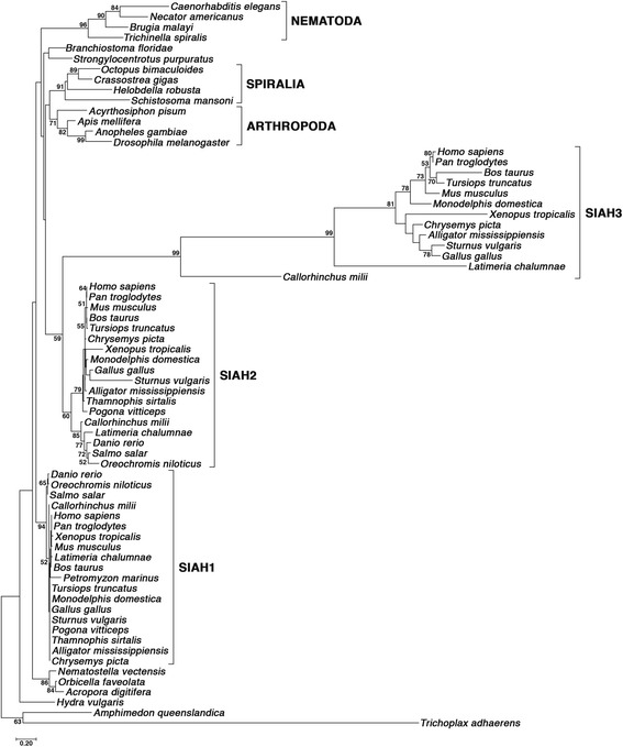
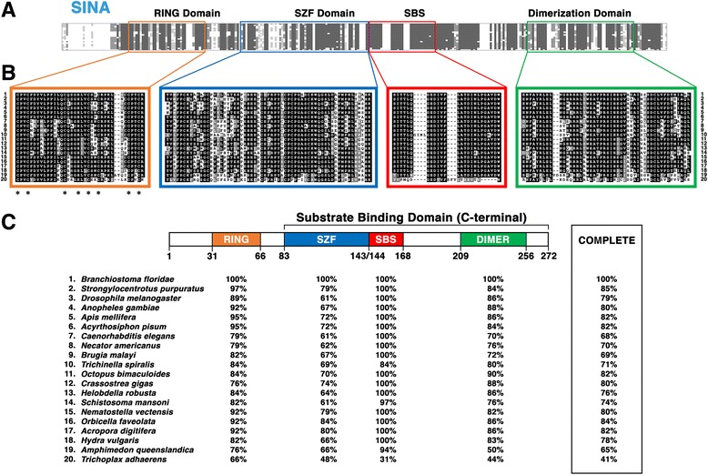
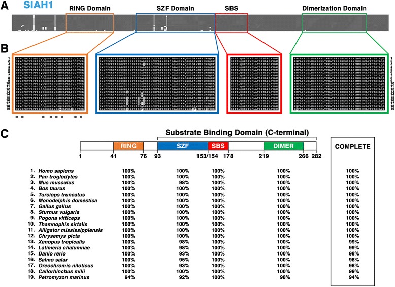
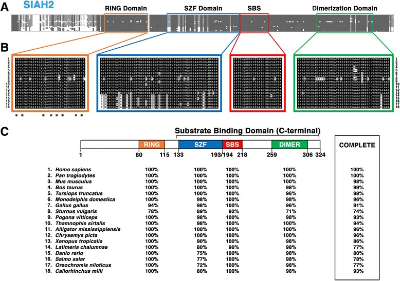
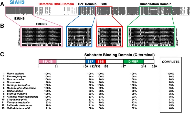
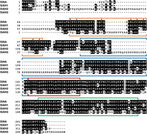
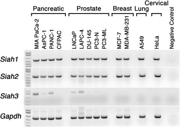
Results: Vertebrate SIAH1, 2, and 3 are the three orthologs to invertebrate SINA protein. SINA and SIAH1 orthologs are found in all major taxa of metazoans. These proteins have four conserved functional domains, known as RING (Really Interesting New Gene), SZF (SIAH-type zinc finger), SBS (substrate binding site) and DIMER (Dimerization). In addition to the siah1 gene, most vertebrates encode two additional siah genes (siah2 and siah3) in their genomes. Vertebrate SIAH2 has a highly divergent and extended N-terminal sequence, while its RING, SZF, SBS and DIMER domains maintain high amino acid identity/similarity to that of SIAH1. But unlike vertebrate SIAH1 and SIAH2, SIAH3 lacks a functional RING domain, suggesting that SIAH3 may be an inactive E3 ligase. The SIAH3 subtree exhibits a high degree of amino acid divergence when compared to the SIAH1 and SIAH2 subtrees. We find that SIAH1 and SIAH2 are expressed in all human epithelial cell lines examined thus far, while SIAH3 is only expressed in a limited subset of cancer cell lines.

Conclusion: Through phylogenetic analyses of metazoan SINA and SIAH E3 ligases, we identified many invariant and divergent amino acid residues, as well as the evolutionarily conserved functional motifs in this medically relevant gene family. Our phylomedicinal study of this unique metazoan SINA/SIAH protein family has provided invaluable evolution-based support towards future effort to design logical, potent, and durable anti-SIAH-based anticancer strategies against oncogenic K-RAS-driven metastatic human cancers. Thus, this method of evolutionary study should be of interest in cancer biology.

Keywords: And conserved functional domains in SINA; Invariant and divergent amino acid residues; Phylogenetic analysis; RAS signal transduction; SIAH1; SIAH2 and SIAH3; SINA/SIAH E3 ligases; Ubiquitin-mediated proteolysis.


Conflict of interest statement

Ethics approval and consent to participate

Not applicable. All metazoan SINA/SIAH sequences are publically available at GenBank and NCBI. Human cancer cell lines were purchased from ATCC. No patient information was used in this study.

Consent for publication

Not applicable.

Competing interests

The authors declare that they have no competing interests.

Publisher’s Note

Springer Nature remains neutral with regard to jurisdictional claims in published maps and institutional affiliations.

Figures

Fig. 1
Fig. 1
Phylogenetic tree of the evolutionarily conserved SINA/SIAH family across metazoan species. The phylogenetic tree was constructed to illustrate the evolutionary relationships of SINA/SIAH family using the representative species from all the major taxa across the entire metazoan kingdom. The LG + G4 + F model was utilized for construction of the tree. The numbers listed on each node represent the bootstrap support value associated with that node after running 100 replicates. All bootstrap values < 50 were eliminated from the tree display. The tree was manually rooted at the node containing the outgroup sequences A. queenslandica and T. adhaerens. Major clades that were recovered by the analysis are indicated by the brackets on the right side
Fig. 2
Fig. 2
Sequence alignment of the invertebrate SINA subfamily reveals its invariant amino acid residues, and the four conserved structural motifs. Sequence comparison of SINA proteins from 20 representative invertebrate species (#1-#20) is shown. a Overview of the entire alignment produced by the 20 invertebrate SINA sequences. Four key functional domains are marked in four distinct colors: RING domain (orange), SZF domain (blue), SBS (red), and DIMER domain (green). b Schematic illustration of amino acid conservation within the 4 domains of the SINA sequences is shown. Amino acid identity is shown as white letters in a black box, amino acid similarity is shown as white letters in a grey box, and amino acid divergence is shown as black letters in a white box. The asterisks located below the RING domain alignment indicate unanimous conservation of the cysteine/histidine zinc-binding residues. c The percentages of amino acid conservation in each distinct domain and the entire SINA sequence between Branchiostoma floridae and each of the representative invertebrate species are shown. The diagram of the domain architecture was based on B. floridae SINA
Fig. 3
Fig. 3
Sequence alignment of the vertebrate SIAH1 subfamily reveals its invariant amino acid residues, and the four conserved structural motifs. Sequence comparison of SIAH1 proteins from 19 representative vertebrate species (#1-#19) is shown. a The level of amino acid conservation in the N-terminal portion (#1 to #40) is high among SIAH1 sequences. Four key functional domains are marked in four distinct colors: RING domain (orange), SZF domain (blue), SBS (red), and DIMER domain (green). b Schematic illustration of amino acid conservation within the 4 domains of the SIAH1 sequences is shown. Amino acid identity is shown as white letters in a black box, amino acid similarity is shown as white letters in a grey box, and amino acid divergence is shown as black letters in a white box. The asterisks located below the RING domain alignment indicate unanimous conservation of the cysteine (Cys)/histidine (His) zinc-binding residues. c The percentages of amino acid conservation in each distinct domain and the entire SIAH1 sequence between human and each of the representative vertebrate species are shown. The diagram of the domain architecture was based on Homo sapiens SIAH1
Fig. 4
Fig. 4
Sequence alignment of the vertebrate SIAH2 subfamily reveals its invariant amino acid residues, and the four conserved structural motifs. Sequence comparison of SIAH2 proteins from 18 representative vertebrate species (#1-#18) is shown. a The level of amino acid divergence in the N-terminal fragments (#1 to #80) is high. Four key functional domains are marked in four distinct colors: RING domain (orange), SZF domain (blue), SBS (red), and DIMER domain (green). b Schematic illustration of amino acid conservation within the 4 domains of the SIAH2 sequences is shown. Amino acid identity is shown as white letters in a black box, amino acid similarity is shown as white letters in a grey box, and amino acid divergence is shown as black letters in a white box. The asterisks located below the RING domain alignment indicate unanimous conservation of the cysteine (Cys)/histidine (His) zinc-binding residues. c The percentages of amino acid conservation in each distinct domain and the entire SIAH2 sequence between human and each of the representative vertebrate species are shown. The diagram of the domain architecture was based on Homo sapiens SIAH2. The SIAH2 sequence for C. milii was incomplete, and these gaps induced by the incompleteness of the sequence were disregarded when calculating conservation across the whole protein
Fig. 5
Fig. 5
Sequence alignment of the vertebrate SIAH3 subfamily reveals its invariant amino acid residues, absence of RING domain, and conserved structural motifs. Sequence comparison of SIAH3 proteins from 13 representative vertebrate species (#1-#13) is shown. a The level of amino acid conservation in the N-terminal fragments is high, while the portion of the protein sequences that would be expected to contain the RING domain is highly divergent compared to SIAH1 and SIAH2. Four key functional domains are marked in four distinct colors: SIAH3 unique N-terminal sequence (S3UNS, pink), SZF domain (blue), SBS (red) and DIMER domain (green). b Schematic illustration of amino acid conservation within the 4 distinct domains of the SIAH3 sequences is shown. Amino acid identity is shown as white letters in a black box, amino acid similarity is shown as white letters in a grey box, and amino acid divergence is shown as black letters in a white box . c The percentages of amino acid conservation in each distinct domain and the entire SIAH3 sequence between human and each of the representative vertebrate species are shown. The diagram of the domain architecture was based on Homo sapiens SIAH3
Fig. 6
Fig. 6
The consensus sequences of SINA, SIAH1, SIAH2, and SIAH3 were aligned to identify the invariant and divergent amino acid residues in this evolutionarily highly conserved SINA/SIAH E3 ligase family. There is a high level of amino acid conservation in the SBD domain in the SINA, SIAH1, SIAH2, and SIAH3 core consensus sequences in the SIAH family. The RING domain is marked by an orange bracket, the SZF domain by a blue bracket, the SBS by a red bracket, and the DIMER domain by a green bracket. Asterisks within the RING domain indicate the position of the invariant cysteine (Cys)/histidine (His) residues in SINA, SIAH1 and SIAH2. Amino acid positions marked with an “X” (instead of a valid one-letter amino acid abbreviation) indicate that the consensus at this site could not be resolved unambiguously. SINA, SIAH1, and SIAH2 share extensive sequence homology between each other in their core consensus sequences, whereas SIAH3 shows dramatic sequence divergence in the corresponding RING and SZF domains to those of SINA, SIAH1, and SIAH2 proteins. SINA, SIAH1, SIAH2, and SIAH3 share high levels of amino acid conservation in the SBS domains
Fig. 7
Fig. 7
SIAH1, SIAH2 and SIAH3 mRNA expression in human cancer cell lines. Semi-quantitative reverse transcription – polymerase chain reaction (RT-PCR) analysis of siah1, siah2 and siah3 mRNA transcript expression in human cancer cell lines is shown. The relative expression levels of siah1, siah2 and siah2 mRNA transcripts in 13 epithelial cancer cells including pancreatic cancer cells (MiaPaCa, AsPC-1, PANC-1 and CFPAC), prostate tumor cell lines (LNCaP, LAPC-4. PC-3-N (normal), PC-3-ML (metastatic)), breast tumor cell lines (MCF-7 and MDA-MB-231), non-small cell lung cancer cell line (A549) and cervical cancer cell line (HeLa) were estimated semi-quantitatively for serial dilutions of the complementary DNA templates. Glyceraldehyde-3-phosphate dehydrogenase (GAPDH) mRNA transcript was used as an internal control. The RT-PCR mixture without cDNA template was used as a negative control

Similar articles

Cited by

References

    1. Greenwald I, Rubin GM. Making a difference: the role of cell-cell interactions in establishing separate identities for equivalent cells. Cell. 1992;68(2):271–281. doi: 10.1016/0092-8674(92)90470-W. - DOI - PubMed
    1. Zipursky SL, Rubin GM. Determination of neuronal cell fate: lessons from the R7 neuron of drosophila. Annu Rev Neurosci. 1994;17:373–397. doi: 10.1146/annurev.ne.17.030194.002105. - DOI - PubMed
    1. Downward J. Targeting RAS signalling pathways in cancer therapy. Nat Rev Cancer. 2003;3(1):11–22. doi: 10.1038/nrc969. - DOI - PubMed
    1. Schubbert S, Shannon K, Bollag G. Hyperactive Ras in developmental disorders and cancer. Nat Rev Cancer. 2007;7:295–308. doi: 10.1038/nrc2109. - DOI - PubMed
    1. Xu C, Liu R, Zhang Q, Chen X, Qian Y, Fang W. The diversification of evolutionarily conserved MAPK cascades correlates with the evolution of fungal species and development of lifestyles. Genome Biol Evol. 2016; - PMC - PubMed

MeSH terms

LinkOut - more resources