Review
. 2017 Oct;42(10):799-811.
doi: 10.1016/j.tibs.2017.07.002. Epub 2017 Aug 4.

How Hsp90 and Cdc37 Lubricate Kinase Molecular Switches

Affiliations
Review

How Hsp90 and Cdc37 Lubricate Kinase Molecular Switches

Kliment A Verba et al. Trends Biochem Sci. 2017 Oct.

Abstract

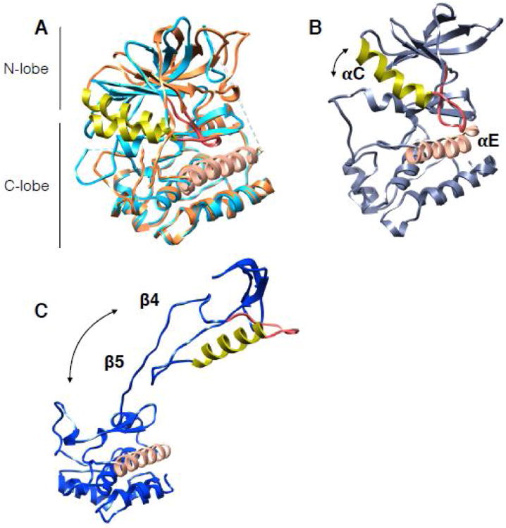
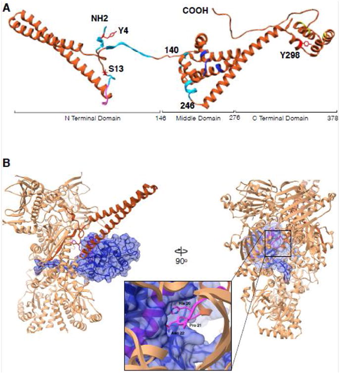
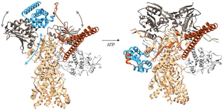
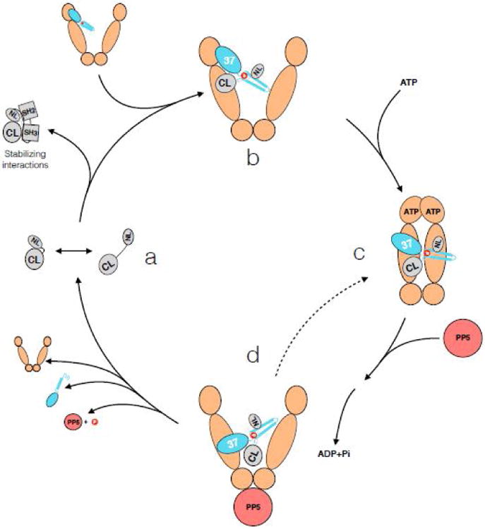
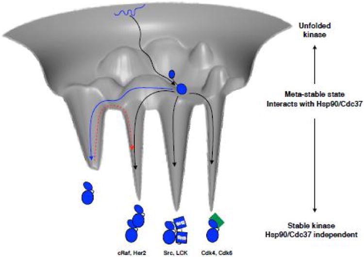
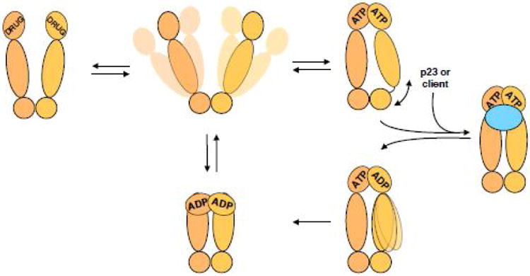
The Hsp90/Cdc37 chaperone system interacts with and supports 60% of the human kinome. Not only are Hsp90 and Cdc37 generally required for initial folding, but many kinases rely on the Hsp90/Cdc37 throughout their lifetimes. A large fraction of these 'client' kinases are key oncoproteins, and their interactions with the Hsp90/Cdc37 machinery are crucial for both their normal and malignant activity. Recently, advances in single-particle cryo-electron microscopy (cryoEM) and biochemical strategies have provided the first key molecular insights into kinase-chaperone interactions. The surprising results suggest a re-evaluation of the role of chaperones in the kinase lifecycle, and suggest that such interactions potentially allow kinases to more rapidly respond to key signals while simultaneously protecting unstable kinases from degradation and suppressing unwanted basal activity.


Figures

Figure 1
Figure 1. Kinase domain architecture and dynamics
The αC helix in all structures is in lime color, adjacent αC-β4 loop is in red. The proteins have been aligned by the αE helix (salmon color helix in C-lobe). Arrows show the direction and relative magnitude of N-lobe motion in relation to C-lobe. (A) Structures of EGFR in the active state (light blue in color, PDB:2ITP) and the inactive state (piper in color, PDB:2GS7). Note the small shift in αC helix between states. (B) Snapshot from 12us all-atom MD simulation on EGFR from Shaw group displaying an opening between the kinase lobes. (C) Cdk4 kinase structure as it is in the cryoEM complex of Hsp90-Cdc37-Cdk4 (PDB:5FWL) showing dramatic unfolding between the kinase lobes and unraveling of β4- β5 strands.
Figure 2
Figure 2. Cdc37 architecture and interactions with Cdk4 kinase in context of Hsp90
(A) Cdc37 structure synthesized from different studies. Residues 1-260 are from cryoEM structure (PDB:5FWL), 261-293 from Hsp90-Cdc37 domain crystal structure (PDB:1US7) and 294-378 from the ensemble solution NMR structure (PDB:2N5X). Cyan color indicates regions interacting with Hsp90 in Hsp90-Cdc37-Cdk4 complex, pink is the kinase interacting loop (HPN motif), blue color are Hsp90 interacting residues as in the crystal structure and in yellow are residues implicated in additional interactions with bRaf kinase by NMR. Y4, S13 and Y298 are known to be phosphorylated and are marked in red. (B) Interactions between Cdc37 and Cdk4 in the context of Hsp90. Only the Cdc37 N terminal domain is depicted for clarity (piper) interacting with the Cdk4 αE helix (pink helix within blue surface) with the loop harboring HPN motif on Cdc37 (pink, zoomed in). Kinase (omitting residues 1-85) depicted as blue transparent surface, threads β4- β5 strands through the lumen of Hsp90 with Hsp90 clamping around them at its middle domain-C terminal domain junction.
Figure 3
Figure 3. Conformational rearrangements of Cdc37 during the Hsp90 cycle
On the left is a modeled complex between Hsp90-Cdc37-Cdk4 where Cdc37-Hsp90 interactions are preserved as in the crystal structure of the fragments (PDB:1US7). The Cdc37 middle domain is colored in blue with the N terminal domain in piper, Hsp90 is in tan with N terminal domains in charcoal and Cdk4 kinase is in light gray. In this state Hsp90's N terminal domains are parted, and the domain of the monomer on the left had to undergo a rotation as to avoid clash between Hsp90's middle domain and Cdc37's middle domain. At this state, we imagine the kinase lobes to be partially open. Upon ATP binding to the Hsp90 N terminal domains, these undergo closure displacing Cdc37 down to the middle domain and stabilizing the kinase in the unfolded state, resulting in the observed cryoEM structure (domain motions are depicted with arrows). Structure on the left corresponds to state (b) in Figure 4 and structure on the right corresponds to state (c) in Figure 4.
Figure 4
Figure 4. Hsp90-kinase cycle
Kinases sample partially open states (CL- C-lobe, NL – N-lobe) (state a). If the kinase spends too much time open at the lobes, for example during early folded state or with a constitutive client, Cdc37 wedges in between the two kinase lobes with its N terminal domain, further unfolding the kinase (state b). Based on NMR data, the C terminus of Cdc37 also makes some kinase interactions at this point. Cdc37 interacts with the N terminal domain of an open Hsp90, utilizing the contacts as in the crystal structure (modeled in Figure 3). Upon binding ATP, Hsp90 undergoes closure, clamping around the unfolded kinase β4-β5 strands, and Cdc37 migrates to the middle domain (state c). Upon ATP hydrolysis by Hsp90 (potentially sequential, with an asymmetric state in between), Hsp90 opens, giving a chance for the kinase to fold (state d). If this was an initial folding interaction for a “non client”, or there is a stabilizing binding partner present nearby for the constitutive client, the stabilized and folded N lobe outcompetes Cdc37 for the C lobe, dissociating the kinase from the complex. However, if there are no stabilizing interactions, Hsp90 may re-bind ATP and enter another cycle (dashed line). This way, the kinase is safely held in a partially folded inactive state, always ready to interact with an appropriate binding partner, checking for its presence with the frequency of Hsp90's ATPase. Dynamic phosphorylation and dephosphorylation by Yes and CKII kinases, and PP5 phosphatase, add a layer of regulation.
Figure 5
Figure 5. Imagined kinase folding free energy landscape
Kinase starts out in an unfolded state (depicted as blue squiggly line at the top of the folding funnel), and likely with the help of general chaperone machinery reaches a partially folded state, perhaps with a rather well folded C-lobe (black arrow to the middle of the folding funnel). From this ensemble of metastable states the kinase interacts with Hsp90-Cdc37. Non-client kinases would then readily access a distinct, well-folded native state and quickly escape the metastable ensemble (blue arrow). If the kinase is a constitutive client, it spends most of its time in the ensemble of partially folded states, without access to a stable state by itself and needs an interacting partner to be stabilized, be it cyclin (green), SH2-SH3 domains or even other kinases (black arrows into deep energy wells). Examples of client kinases representing each mode of stabilization are given below the cartoons. Non-client kinases would have to pay a considerable energy penalty to transition out of the energetically deep native state into an effector bound state (red dashed arrow). Hsp90 client kinases however, being in a structurally uncommitted state are always ready to interact with effectors, foregoing the need to pay the energy penalty. Importantly, Hsp90-Cdc37 may also catalyze the transitions between interactions with different effectors.
Figure I
Figure I. Hsp90's ATPase cycle
Hsp90 dimer (shades of tan) samples a variety of open states without a nucleotide. Upon binding of ATP, Hsp90 adopts a closed state with the N terminal domains rotating and dimerizing. Without additional binding partners such closed state is asymmetric at the interface between Hsp90's middle and C terminal domains. However, binding of co-chaperones like p23 or co-chaperones with clients, like Cdc37-kinase (either case is shown as blue oval) causes Hsp90 to adopt a symmetric state. ATP is then hydrolyzed in the N terminal domains, potentially sequentially with a switch in the asymmetry between the Hsp90 monomers, on the way to a more compact ADP state. Release of the nucleotide returns Hsp90 back to the equilibrium of open states. Inhibitors, like geldanamycin, 17AAG, radicicol, etc break the ATPase cycle of Hsp90, stalling it in the more compact state, yet different from the ATP bound state.

References

    1. Taylor SS, Keshwani MM, Steichen JM, Kornev AP. Evolution of the eukaryotic protein kinases as dynamic molecular switches. Philos Trans R Soc Lond B Biol Sci. 2012;367:2517–2528. - PMC - PubMed
    1. Endicott JA, Noble ME, Johnson LN. The structural basis for control of eukaryotic protein kinases. Annu Rev Biochem. 2012;81:587–613. - PubMed
    1. Zhang J, Yang PL, Gray NS. Targeting cancer with small molecule kinase inhibitors. Nat Rev Cancer. 2009;9:28–39. - PubMed
    1. Brugge J, Yonemoto W, Darrow D. Interaction between the Rous sarcoma virus transforming protein and two cellular phosphoproteins: analysis of the turnover and distribution of this complex. Mol Cell Biol. 1983;3:9–19. - PMC - PubMed
    1. Gierasch LM, Horwich A, Slingsby C, Wickner S, Agard D. Series in structural biology Vol 6. World Scientific; New Jersey: 2016. 1 online resource (vii, 319 pages)

MeSH terms