. 2017 Nov 1;25(11):2490-2501.
doi: 10.1016/j.ymthe.2017.06.016. Epub 2017 Aug 4.

Adipose Stem Cell Therapy Mitigates Chronic Pancreatitis via Differentiation into Acinar-like Cells in Mice

Affiliations

Adipose Stem Cell Therapy Mitigates Chronic Pancreatitis via Differentiation into Acinar-like Cells in Mice

Zhen Sun et al. Mol Ther. .

Abstract

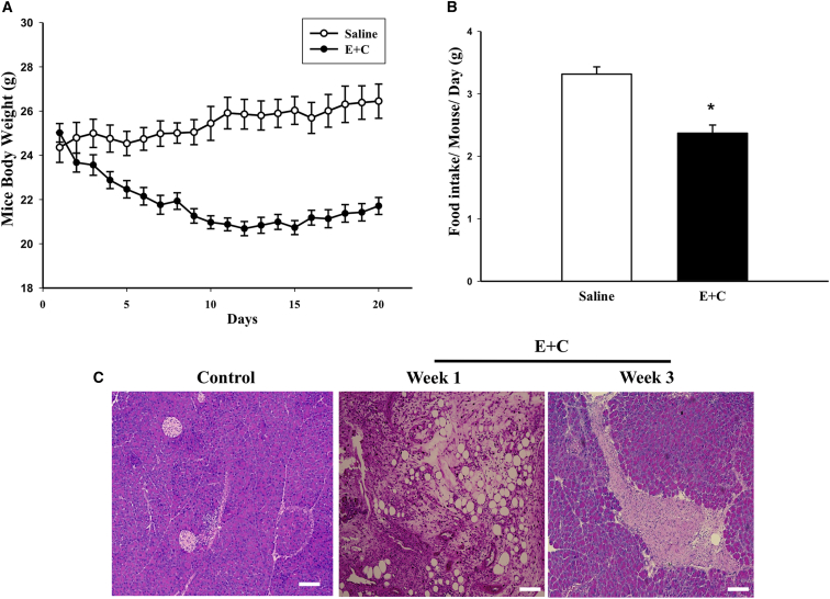
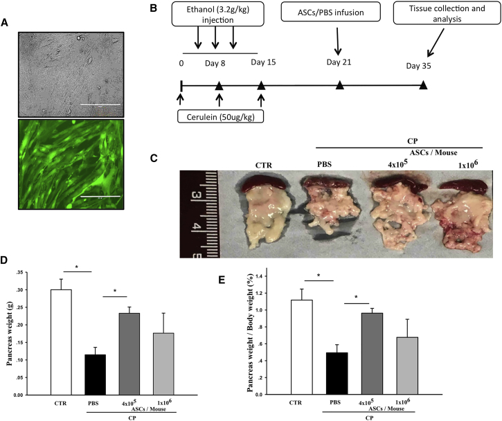
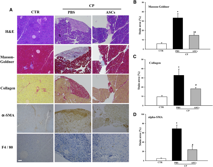
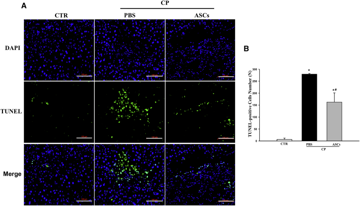
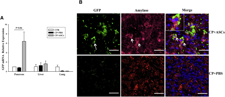
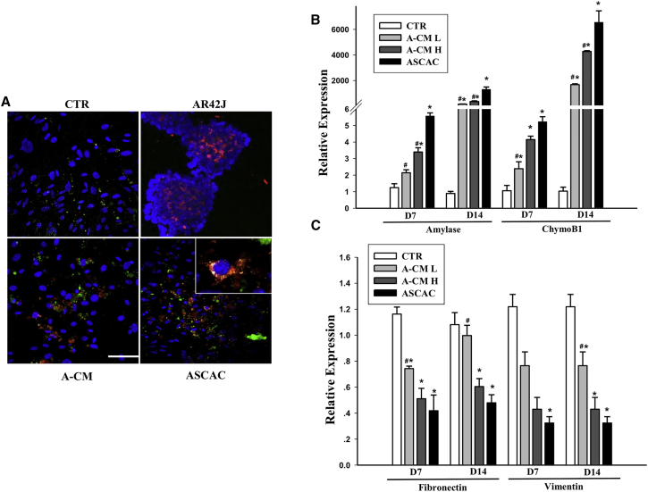
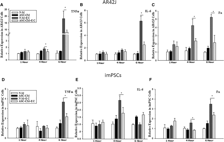
The objective of this study was to assess the capacity of adipose-derived mesenchymal stem cells (ASCs) to mitigate disease progression in an experimental chronic pancreatitis mouse model. Chronic pancreatitis (CP) was induced in C57BL/6 mice by repeated ethanol and cerulein injection, and mice were then infused with 4 × 10⁵ or 1 × 10⁶ GFP+ ASCs. Pancreas morphology, fibrosis, inflammation, and presence of GFP+ ASCs in pancreases were assessed 2 weeks after treatment. We found that ASC infusion attenuated pancreatic damage, preserved pancreas morphology, and reduced pancreatic fibrosis and cell death. GFP+ ASCs migrated to pancreas and differentiated into amylase+ cells. In further confirmation of the plasticity of ASCs, ASCs co-cultured with acinar cells in a Transwell system differentiated into amylase+ cells with increased expression of acinar cell-specific genes including amylase and chymoB1. Furthermore, culture of acinar or pancreatic stellate cell lines in ASC-conditioned medium attenuated ethanol and cerulein-induced pro-inflammatory cytokine production in vitro. Our data show that a single intravenous injection of ASCs ameliorated CP progression, likely by directly differentiating into acinar-like cells and by suppressing inflammation, fibrosis, and pancreatic tissue damage. These results suggest that ASC cell therapy has the potential to be a valuable treatment for patients with pancreatitis.

Keywords: acinar cells; adipose stem cell therapy; differentiation; ethanol and cerulean-induced chronic pancreatitis mouse model; fibrosis; inflammation; mesenchymal stem cells.


Figures

Figure 1
Figure 1
Ethanol and Cerulean-Induced CP Mouse Model (A) Body weight changes in mice after saline (CTR) or ethanol and cerulein (E+C) injections (n = 10 per group). (B) Food intake in mice treated with saline or ethanol and cerulein 2 weeks after CP induction. (C) Representative H&E staining of pancreatic tissue from mice receiving saline (control) or ethanol and cerulein 1 and 3 weeks after injection. Scale bars, 100 μm. Data presented are mean ± SEM. Differences were compared by one-way ANOVA; *p < 0.05.
Figure 2
Figure 2
Characteristics of CP Mice (A) Micrographs of GFP+ ASCs observed by light or fluorescent microscopy. Scale bars, 100 μm. (B) Schematic diagram of CP induction, cell infusion, and tissue collection and analysis. (C) Images of pancreases collected from normal control, CP control, or CP mice treated with ASCs (4 × 105 or 1 × 106 cells/mouse). (D and E) Pancreas weights (D) and percent ratio of pancreas weight to body weight (E) in control, CP control, or CP mice treated with ASCs (4 × 105 or 1 × 106 cells/mouse). Data presented are mean ± SEM. Differences were compared by one-way ANOVA; *p < 0.05.
Figure 3
Figure 3
Effects of ASC Injection on Pancreas Damage, Fibrosis, and Macrophage Infiltration (A) Histological analysis of pancreas tissue sections from normal control or CP mice treated either with PBS or ASCs at 4 × 105 cells/mouse. H&E staining, Masson-Goldner-trichrome staining, and staining with antibodies against collagen, α-SMA, and F4/80 are shown. Scale bar, 100 μm. (B–D) Percentages of positive area in total pancreatic area of (B) Masson-Goldner, (C) collagen, and (D) α-SMA. Data are means ± SEMs. *p < 0.05 compared with normal control; #p < 0.05 compared with CP-PBS group, one-way ANOVA test.
Figure 4
Figure 4
ASC Infusion Protects against Pancreatic Cell Death (A) Immunostaining of TUNEL+ cells in the pancreas from normal control, CP mice treated with PBS, or ASCs (4 × 105 cells/mouse) 2 weeks after infusion. Scale bars, 100 μm. (B) Average numbers of TUNEL+ cells in pancreas sections. Data presented are mean ± SEM. Differences were compared by one-way ANOVA; *p < 0.05 compared with normal control; #p < 0.05 compared with CP-PBS group.
Figure 5
Figure 5
Differentiation of Infused ASCs into Amylase+ and Insulin+ Cells in the Treated Pancreas (A) Real-time RT-PCR analysis of mRNA expression of GFP in the pancreas, liver, and lungs from normal controls, CP controls, and ASC-treated CP mice 2 weeks after injection. (B) Immunohistochemical staining of pancreatic tissues from mice receiving ASCs (upper panels) or PBS (lower panels) with the anti-GFP and the anti-amylase antibodies. Green represents GFP+ cells, and red identifies amylase+ cells. Nuclei are stained blue (DAPI). Scale bar, 50 μm. Data presented are mean ± SEM. Differences were compared by one-way ANOVA.
Figure 6
Figure 6
ASCs Differentiate into Amylase+ Cells In Vitro When Co-cultured with Acinar Cells or in Acinar Cell-Conditioned Medium (A) ASCs cultured alone (CTR), cultured in AR42J-conditioned medium (A-CM), or co-cultured with AR42J cells (ASCAC) were stained for GFP (green) and amylase (red). AR42J cells were used as a positive control for amylase staining. Nuclei are stained blue (DAPI). Scale bar, 50 μm. (B and C) Relative expression of amylase 1 and chymoB1 (B) and fibronectin and vimentin (C) after 7 or 14 days of culture, in ASCs cultured alone (CTR), in ASCs cultured in low-dose AR42J-conditioned medium (A-CM-L), ASCs cultured in high-dose AR42J-conditioned medium (A-CM-H), or ASCs co-cultured with acinar cells. Data presented are mean ± SEM. Differences were compared by one-way ANOVA; *p < 0.05 versus CTR, #p < 0.05 versus ASCAC.
Figure 7
Figure 7
Co-culture with ASC-Conditioned Medium Inhibits Ethanol and Cerulein-Induced Expression of Proinflammatory Cytokines in AR42J Cells and imPSCs (A–F) Expression of TNF-α (A and D), IL-6 (B and E), and fibronectin (C and F) in AR42J cells or imPSCs. Data are representative of at least three individual experiments. Data presented are mean ± SEM. Differences were compared by one-way ANOVA; *p < 0.05. ASC-CM, cells cultured in ASC-conditioned medium; ASC-CM+EC, cells cultured in ASC-conditioned medium challenged with ethanol and cerulein; N-M, cells cultured in DMEM/F12; N-M+EC, cells cultured in DMEM/F12 challenged with ethanol and cerulein.

Comment in

  • Adipose Stem Cell Therapy for Chronic Pancreatitis.
    Ben Nasr M, Frumento D, Fiorina P. Ben Nasr M, et al. Mol Ther. 2017 Nov 1;25(11):2438-2439. doi: 10.1016/j.ymthe.2017.10.007. Epub 2017 Oct 19. Mol Ther. 2017. PMID: 29055621 Free PMC article. No abstract available.

References

    1. Lankisch P.G. Natural course of chronic pancreatitis. Pancreatology. 2001;1:3–14. - PubMed
    1. Adams D.B., Davis B.R., Anderson M.C. Colonic complications of pancreatitis. Am. Surg. 1994;60:44–49. - PubMed
    1. Etemad B., Whitcomb D.C. Chronic pancreatitis: diagnosis, classification, and new genetic developments. Gastroenterology. 2001;120:682–707. - PubMed
    1. Tieftrunk E., Demir I.E., Simon P., Friess H., Ceyhan G.O. Evidence of pancreatic neuropathy and neuropathic pain in hereditary chronic pancreatitis. Pancreatology. 2013;13:629–630. - PubMed
    1. Marks I.N., Bank S. The aetiology, clinical features and diagnosis of pancreatitis in the South Western Cape; a review of 243 cases. S. Afr. Med. J. 1963;37:1039–1053. - PubMed

MeSH terms

LinkOut - more resources