. 2017 Aug 22;114(34):9080-9085.
doi: 10.1073/pnas.1708448114. Epub 2017 Aug 7.

Identification of the Tau phosphorylation pattern that drives its aggregation

Affiliations

Identification of the Tau phosphorylation pattern that drives its aggregation

Clément Despres et al. Proc Natl Acad Sci U S A. .

Abstract

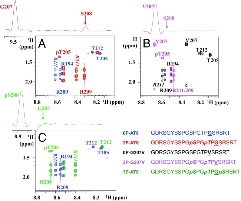
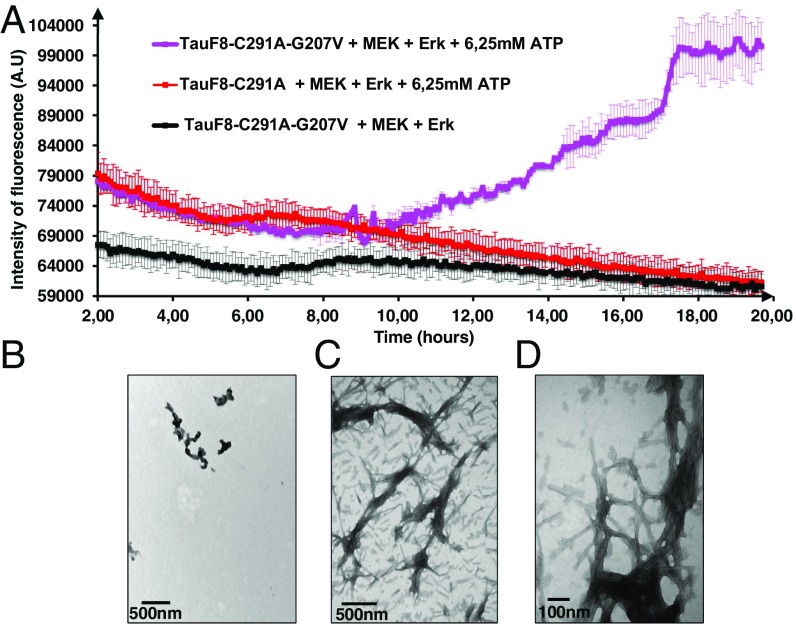
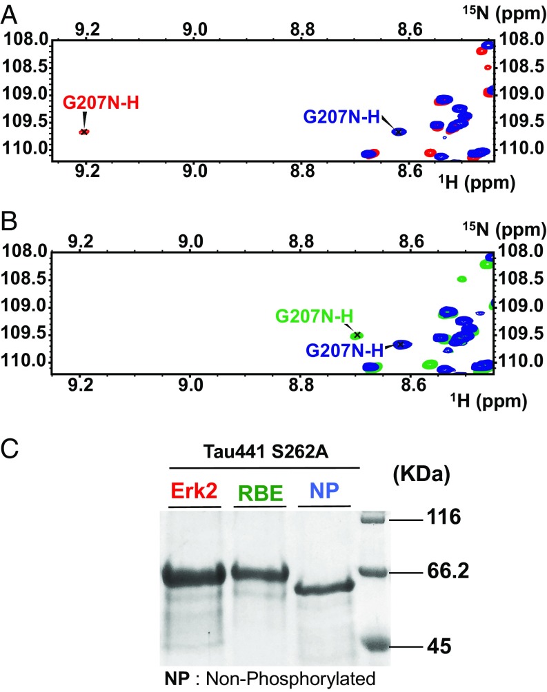
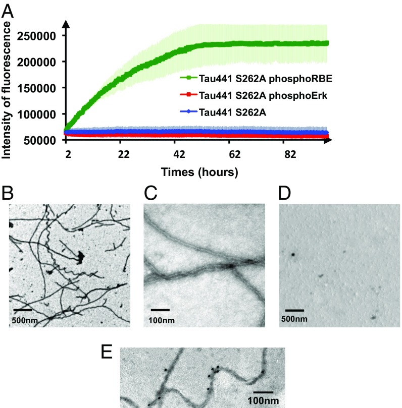
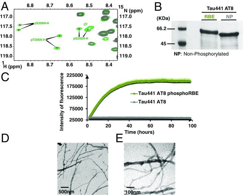
Determining the functional relationship between Tau phosphorylation and aggregation has proven a challenge owing to the multiple potential phosphorylation sites and their clustering in the Tau sequence. We use here in vitro kinase assays combined with NMR spectroscopy as an analytical tool to generate well-characterized phosphorylated Tau samples and show that the combined phosphorylation at the Ser202/Thr205/Ser208 sites, together with absence of phosphorylation at the Ser262 site, yields a Tau sample that readily forms fibers, as observed by thioflavin T fluorescence and electron microscopy. On the basis of conformational analysis of synthetic phosphorylated peptides, we show that aggregation of the samples correlates with destabilization of the turn-like structure defined by phosphorylation of Ser202/Thr205.

Keywords: Alzheimer’s disease; NMR; Tau; aggregation; phosphorylation.


Conflict of interest statement

The authors declare no conflict of interest.

Figures

Fig. 1.
Fig. 1.
Breaking the turn-like structure by the Gly207Val mutation or by phosphorylation of Ser208. Zooms of the homonuclear TOCSY spectra of the nonmodified and phosphorylated peptides centered on the Thr205 and Arg209/211 residues. (Upper) The 1D traces are extracted from the corresponding NOESY spectra on the phosphorylated peptides. (A) Phosphorylation of the WT peptide at Ser202/Thr205, as in the 2P-AT8 peptide, leads to an important shift for the Arg209 amide proton and is accompanied by an NOE contact between the amide protons of Gly207 and Ser208, indicative of the phosphorylation-induced turn (14). (B) When Gly207 is substituted for a Val, the absence of an NOE contact in the 2P-G207V peptide between the amide protons of Val207 and Ser208 and the reduced shift for Arg209 indicate a destabilization of the turn-like structure. (C) Additional phosphorylation of Ser208 in the 3P-AT8 peptide has a similar effect as the Gly-to-Val substitution at position 207.
Fig. 2.
Fig. 2.
Destabilizing the turn by the G207V mutation combined with ERK2-mediated phosphorylation leads to aggregation of the TauF8 fragment. (A) Aggregation of TauF8 (without external inducers) during the phosphorylation reaction, as followed by ThT emission at 490 nm. An increase in ThT emission is observed only for the G207V mutant. Error bars correspond to three independent aggregation experiments, with new batches of TauF8, TauF8-G207V, MEK, and ERK2 kinases. (BD) TEM at the end point of the phosphorylation reactions shows a large amount of fibrils after phosphorylation of the TauF8-G207V mutant (B and C), but only a smattering of fibril-like structures for the wt TauF8 (D).
Fig. 3.
Fig. 3.
Phosphorylation of Ser208 by RBE, but not ERK2, kinase activity promotes aggregation of Tau441-S262A. (A and B) Glycine region from the 1H-15N HSQC spectra of Tau441-S262A (blue), Tau441-S262A phosphorylated by ERK2 (red), and Tau441-S262A phosphorylated by RBE (green). The downfield shift for the G207 amide proton resonance observed in the ERK2-phosphorylated sample is absent after phosphorylation with RBE. (C) SDS/PAGE analysis of Tau441-S262A before and after phosphorylation by ERK2 and RBE. By comparison with the native protein (lane NP), a similar gel shift is observed for the two phosphorylated samples. The pS208 and pS356 sites phosphorylated solely by the RBE cannot be distinguished by SDS/PAGE.
Fig. 4.
Fig. 4.
Additional phosphorylation of Ser208 by RBE promotes aggregation of Tau441-Ser262A (A) Aggregation of Tau441-S262A (blue), Tau441-S262A phosphorylated by ERK2 (red), and Tau441-S262A phosphorylated by RBE (green) followed by ThT emission at 490 nm. An increase in ThT emission at 490 nm is observed only for the 3P-AT8 Tau441-S262A protein. Error bars correspond to three independent aggregation experiments, with new batches of proteins and different RBEs. (B–D) TEM images at the end point of the aggregation assay of Tau441-S262A phosphorylated by RBE (B and C) or by Erk (D) confirm the results obtained in the aggregation assay. Large amounts of fibrils are observed only for Tau441-S262A phosphorylated by RBE. (E) Immunogold electron microscopy of the fibers obtained with Tau441-S262A phosphorylated by RBE. AT8 indeed stains the fibers in a similar manner as AD brain-derived fibers.
Fig. 5.
Fig. 5.
The 3P-AT8 motif alone is sufficient to drive aggregation. (A) Region of the 1H-15N HSQC spectrum of RBE phosphorylated Tau441-AT8. Because of incomplete phosphorylation, two resonances for each phospho-site are identified. (B) SDS/PAGE analysis of Tau441-AT8 before and after phosphorylation by RBE. Whereas a gel shift is still visible on phosphorylation, it is less pronounced in this mutant, where most phosphorylation sites have been removed. (C) Aggregation of RBE phosphorylated Tau441-AT8 as followed by ThT emission at 490 nm confirms phosphorylation of the Ser202/Thr205/Ser208 epitope is sufficient to trigger aggregation. (D and E) TEM images at the end point of the aggregation reveal a network of fibers when RBE-phosphorylated Tau-AT8 is allowed to aggregate for 4 d. Error bars correspond to two independent experiments with the same Tau441-AT8 batch, but with different RBEs.

References

    1. Goedert M, Spillantini MG. A century of Alzheimer’s disease. Science. 2006;314:777–781. - PubMed
    1. Glenner GG, Wong CW. Alzheimer’s disease: Initial report of the purification and characterization of a novel cerebrovascular amyloid protein. Biochem Biophys Res Commun. 1984;120:885–890. - PubMed
    1. Kosik KS, Joachim CL, Selkoe DJ. Microtubule-associated protein tau (tau) is a major antigenic component of paired helical filaments in Alzheimer disease. Proc Natl Acad Sci USA. 1986;83:4044–4048. - PMC - PubMed
    1. Grundke-Iqbal I, et al. Microtubule-associated protein tau. A component of Alzheimer paired helical filaments. J Biol Chem. 1986;261:6084–6089. - PubMed
    1. Wood JG, Mirra SS, Pollock NJ, Binder LI. Neurofibrillary tangles of Alzheimer disease share antigenic determinants with the axonal microtubule-associated protein tau (tau) Proc Natl Acad Sci USA. 1986;83:4040–4043. - PMC - PubMed

Publication types

MeSH terms

LinkOut - more resources