. 2017 Sep 15;199(6):2030-2042.
doi: 10.4049/jimmunol.1602183. Epub 2017 Aug 7.

α3-Deletion Isoform of HLA-A11 Modulates Cytotoxicity of NK Cells: Correlations with HIV-1 Infection of Cells

Affiliations

α3-Deletion Isoform of HLA-A11 Modulates Cytotoxicity of NK Cells: Correlations with HIV-1 Infection of Cells

Xi-He Zhang et al. J Immunol. .

Abstract

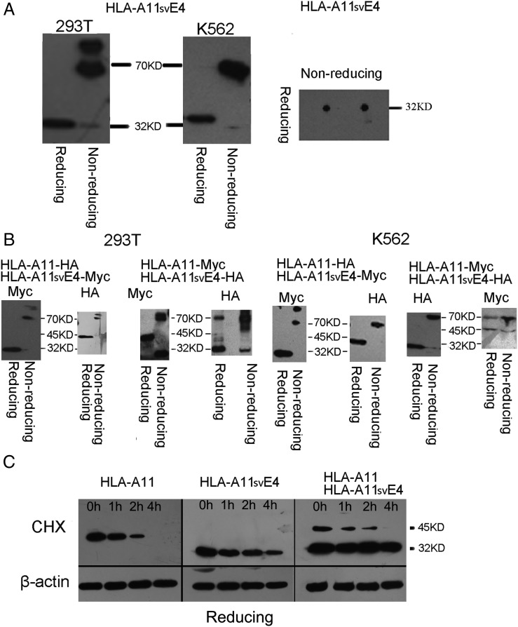
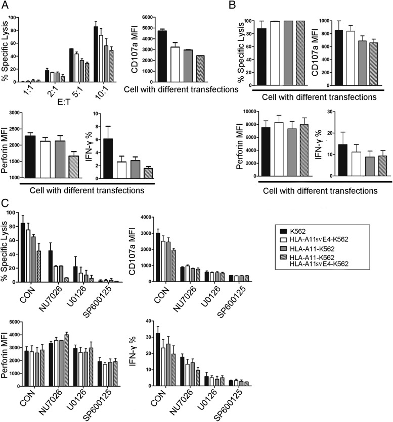
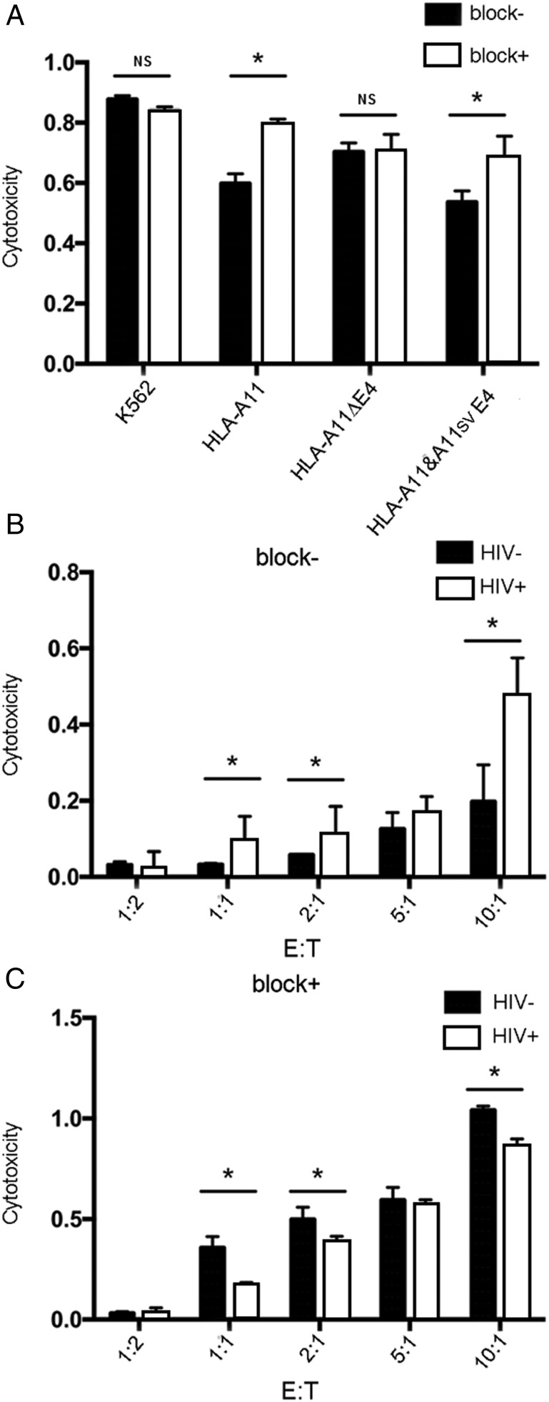
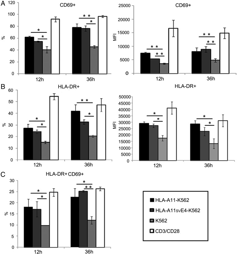
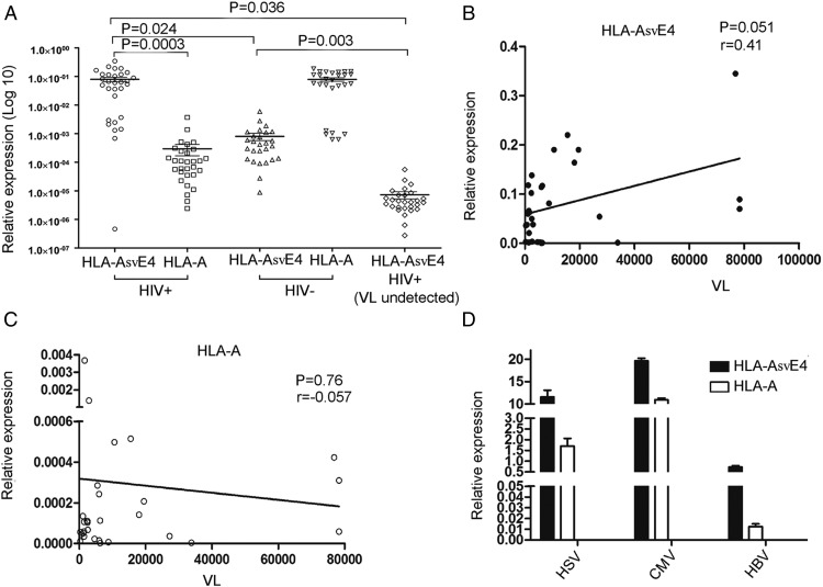
Alternative splicing occurs frequently in many genes, especially those involved in immunity. Unfortunately, the functions of many alternatively spliced molecules from immunologically relevant genes remain unknown. Classical HLA-I molecules are expressed on almost all nucleated cells and play a pivotal role in both innate and adaptive immunity. Although splice variants of HLA-I genes have been reported, the details of their functions have not been reported. In the current study, we determined the characteristics, expression, and function of a novel splice variant of HLA-A11 named HLA-A11svE4 HLA-A11svE4 is located on the cell surface without β2-microglobulin (β2m). Additionally, HLA-A11svE4 forms homodimers as well as heterodimers with HLA-A open conformers, instead of combining with β2m. Moreover, HLA-A11svE4 inhibits the activation of NK cells to protect target cells. Compared with β2m and HLA-A11, the heterodimer of HLA-A11svE4 and HLA-A11 protected target cells from lysis by NK cells more effectively. Furthermore, HLA-AsvE4 expression was upregulated by HIV-1 in vivo and by HSV, CMV, and hepatitis B virus in vitro. In addition, our findings indicated that HLA-A11svE4 molecules were functional in activating CD8+ T cells through Ag presentation. Taken together, these results suggested that HLA-A11svE4 can homodimerize and form a novel heterodimeric complex with HLA-A11 open conformers. Furthermore, the data are consistent with HLA-A11svE4 playing a role in the immune escape of HIV-1.


Figures

FIGURE 1.
FIGURE 1.
Identification of a novel, alternatively spliced transcript of HLA-A11 that encodes a truncated protein. (A) Representation of normal splicing: all seven introns are excised and all eight exons are retained. This form of mRNA corresponds to an ∼1.2-kb band on agarose gel electrophoresis. (B) Representative novel alternative HLA-A transcript wherein exon 4 is deleted together with seven introns and exons 1–3 and 5–8 are retained integrally (verified by sequencing analysis). This form of mRNA corresponds to an ∼0.8-kb band on agarose gel electrophoresis. Exons are represented as boxes; introns are represented by thick lines.
FIGURE 2.
FIGURE 2.
Distribution and expression of HLA-A11 and HLA-A11svE4 proteins in transfected K562 and 293 T cells. (A) The cDNAs encoding HLA-A11 and HLA-A11svE4 were subcloned into pTomo which have a GFP tag and pEGFP to generate C-terminal fusions of HLA-A11 and HLA-A11svE4 with the two vectors; K562 and 293T cells were transfected with these plasmids. Stably expressing K562 and transiently expressing 293T cells were visualized using laser confocal microscopy and images were examined at 60× objective lens magnification. (B) Surface expression of β2m molecules on stable K562 transfectants which expressed either HLA-A11 and/or HLA-A11svE4 was detected by flow cytometry using a FITC-conjugated monoclonal anti-β2m (clone 2M2) Ab. (C) Representative immunoblotting experiment demonstrating the expression of HLA-A11 and HLA-A11svE4 proteins in PBMCs and K562 cells stably transfected with the HLA-A11 and HLA-A11svE4 transcript.
FIGURE 3.
FIGURE 3.
HLA-A11svE4 isoforms homodimerize and heterodimerize with full-length HLA-A11 and are more stable than heterodimers of HLA-A11 and β2m. (A) Monomers and homodimers of HLA-A11svE4 isoforms were identified from transfected cell lysates by Western blotting using reducing and nonreducing two-dimensional SDS-PAGE. Right panel, two-dimensional SDS-PAGE analysis of HLA-A11svE4 homodimerization. Lysates of 293T and K562 cells were analyzed by two-dimensional SDS-PAGE (first dimension, nonreducing; second dimension, reducing). (B) HLA-A11svE4 associates with HLA-A11. HLA-A11 and HLA-A11svE4 were labeled with either HA or myc tags. Plasmids containing these cDNAs were cotransfected into K562 and 293T cells. Lysates of these cells coexpressing myc- and HA-tagged HLA-A11 and HLA-A11svE4 were immunoblotted with anti-HA and anti-myc mAbs. (C) K562 cells stably transfected with HA–HLA-A11svE4 and/or HA–HLA-A11 were divided into four equal parts which were treated with CHX (25 μg/ml) for 0, 1, 2, or 4 h. β-actin served as a loading control. For HA-HLA-A11–HA-HLA-A11svE4 cotransfected cells, lysates were electrophoresed under nonreducing and reducing conditions.
FIGURE 4.
FIGURE 4.
Expression of HLA-A11 and HLA-A11svE4 in different cohorts of PBMCs stimulated by mitogens. (A) B, NK, CD8+, CD4+ T cells, and monocytes were selected by microbeads from PBMCs expressing HLA-A11. RNA from each lymphocyte subpopulation and PBMCs (control) were extracted and then qRT-PCR was performed. The relative expressions of HLA-A11 and HLA-A11svE4 from each subpopulation were compared with that of PBMCs. (B) PBMCs were incubated with Con A, PHA, SPA, LPS, and PWM in serial concentration gradients for 24 h at 37°C. The most effective concentration of each mitogen is shown (10 μg/ml Con A, 10 μg/ml PHA, 10 μg/ml SPA, 10 ng/ml LPS, and 1 μg/ml PWM). RNA from each lymphocyte group and PBMCs were extracted and subjected to qRT-PCR. The relative expressions of HLA-A11 and HLA-A11svE4 in each group were compared with that of PBMCs. Each experiment was performed independently three times.
FIGURE 5.
FIGURE 5.
NK cell cytotoxicity and cytokine release are inhibited by HLA-A11svE4. (A) NK cell cytotoxicity assays and cytokine release with HLA-A11 and HLA-A11svE4 transfectants. The results are expressed as the percentage of lysis recorded after a 6-h lactate dehydrogenase release assay. The E:T ratios were 1:1, 2:1, 5:1, and 10:1. CD107a, IFN-γ, and perforin release were measured by flow cytometry using corresponding mAbs. (B) Agonists of KIR3DL2 induced cytotoxic lysis of HLA-A11–, HLA-A11svE4–, or HLA-A11– and HLA-A11svE4–expressing target cells by NK cells. NK cells were pretreated with an anti-KIR3DL2 mAb (10 μg/ml) before incubation with target cells at an E:T ratio of 10:1. (C) Inhibition of MAPK and DNA-PKcs pathways prevents NK cell cytotoxic lysis of HLA-A11, HLA-A11svE4, or HLA-A11–HLA-A11svE4 transfectants. NK cells were pretreated with different doses of MAPK inhibitors of JNK (SP600125; 50 μM), MEK (U0126; 20 μM), or Akt inhibitor VIII (NU7026; 20 μM) for 2 h at 37°C. Each experiment was performed independently three times.
FIGURE 6.
FIGURE 6.
HLA-A11svE4 inhibits NK lysis of HIV+ CD4+ T cell. (A) NK cell cytotoxicity assays with HLA-A11 and HLA-A11svE4 transfectants. Both NK cells and K562 cells were divided to two cohorts: one of them was incubated with the leaf Ab to block HLA-A11 and the other was used as the control. (B and C) NK and CD4+ T cells were separated from same PBMC sample and CD4+ T cells were infected with HIV-1 for 24 h. NK and HIV+ CD4+ T cells were coincubated with different ratios: 1:2, 1:1, 2:1, 5:1, and 10:1; and anti-HLA Abs were added (C) and another cohort acted as the control (B). Each experiment was performed independently three times. Bars, mean with SD. *p < 0.05, unpaired two-tailed t test.
FIGURE 7.
FIGURE 7.
HLA-A11svE4 activates CD8+ T cells by presenting Ags. K562 cells transfected with either the vector alone, HLA-A11, or HLA-A11svE4 were incubated with 1 μg/ml of LMP-2(340–349) (SSCSSCPLSK, HLA-A11) at 37°C for 2 h and then washed twice. CD8+ T cells were isolated negatively from fresh PBMCs and 2 × 10⁵ CD8+ T cells were incubated with peptide-pulsed K562 cells at ratio of 25:1 containing CD28 mAb. CD8+ T cells with CD3/CD28 beads (bead/cell ratio of 1:1) were set as positive control. Cells were collected at 12 and 36 h, and the expressions of CD8, CD69, and HLA-DR were measured by flow cytometry using corresponding mAbs. (A) Percentage of CD8+ HLA-DR+ T cells and MFI of HLA-DR; (B) Percentage of CD8+ CD69+ T cells and MFI of CD69; (C) Percentage of CD8+, HLA-DR+, and CD69+ T cells. Each experiment was performed independently three times. Bars, mean with SD. *p < 0.05, **p < 0.01, unpaired two-tailed t test.
FIGURE 8.
FIGURE 8.
Expression of HLA-A and HLA-AsvE4 in HIV+ and HIV cohorts and their relationship with HIV viral load. (A) The whole blood of 30 HIV-1+ individuals not taking a mixture of HIV drugs, 27 HIV-1 individuals, and 28 individuals treated with a mixture of HIV drugs and without detectable levels of HIV virus were collected. mRNA levels of HLA-A and HLA-AsvE4 of the three groups were examined by qRT-PCR, and viral load was measured using kits. Relative expression of HLA-A and HLA-AsvE4 was normalized to β-actin expression. (B and C) The relationship of HLA-A and HLA-AsvE4 with HIV viral load, respectively. (D) HSV, HBV, and CMV upregulated HLA-AsvE4 in vitro. The RNA of MRC5 cells (with or without CMV), vero cells (with or without HSV), and Hep and Hep2.215 (HBV+) were extracted, and qRT-PCR was performed to detect HLA-A and HLA-AsvE4. Relative expression of HLA-A and HLA-AΔE4 was normalized to β-actin expression. Expression of HLA-A and HLA-AsvE4 in CMV-, HBV-, and HSV-infected cell lines was compared with that in cells without CMV, HBV, and HSV. Each experiment was performed independently three times. Bars, mean with SD.
FIGURE 9.
FIGURE 9.
HLA-A11SVE4 prohibits HIV elimination. (A) Normal cells present HLA-I, and when NK cells interact with these cells, their activation is inhibited. (B) In HIV-1–infected cells where HLA-I was downregulated, NK cells became activated, leading to lysis of virally infected cells to prevent virus duplication. (C) HIV-1 downregulates full-length HLA and upregulates HLA-AsvE4, which interacts with KIR and inhibits NK cell activation, thereby protecting infected cells.

Similar articles

Cited by

References

    1. Raghavan S., Selvaraj P., Swaminathan S., Narendran G. 2009. Short communication: association of HLA-A*1101 with resistance and B*4006 with susceptibility to HIV and HIV-TB: an in silico analysis of promiscuous T cell epitopes. AIDS Res. Hum. Retroviruses 25: 1023–1028. - PubMed
    1. Li L., Bouvier M. 2004. Structures of HLA-A*1101 complexed with immunodominant nonamer and decamer HIV-1 epitopes clearly reveal the presence of a middle, secondary anchor residue. J. Immunol. 172: 6175–6184. - PubMed
    1. Harrer T., Harrer E., Barbosa P., Kaufmann F., Wagner R., Brüggemann S., Kalden J. R., Feinberg M., Johnson R. P., Buchbinder S., Walker B. D. 1998. Recognition of two overlapping CTL epitopes in HIV-1 p17 by CTL from a long-term nonprogressing HIV-1-infected individual. J. Immunol. 161: 4875–4881. - PubMed
    1. Beyrer C., Artenstein A. W., Rugpao S., Stephens H., VanCott T. C., Robb M. L., Rinkaew M., Birx D. L., Khamboonruang C., Zimmerman P. A., et al. Chiang Mai HEPS Working Group 1999. Epidemiologic and biologic characterization of a cohort of human immunodeficiency virus type 1 highly exposed, persistently seronegative female sex workers in northern Thailand. J. Infect. Dis. 179: 59–67. - PubMed
    1. Sriwanthana B., Hodge T., Mastro T. D., Dezzutti C. S., Bond K., Stephens H. A., Kostrikis L. G., Limpakarnjanarat K., Young N. L., Qari S. H., et al. 2001. HIV-specific cytotoxic T lymphocytes, HLA-A11, and chemokine-related factors may act synergistically to determine HIV resistance in CCR5 delta32-negative female sex workers in Chiang Rai, northern Thailand. AIDS Res. Hum. Retroviruses 17: 719–734. - PubMed

Publication types