

A tip from the nose: rhinocerebral mucormycosis in a patient with alcoholic liver cirrhosis and cocaine abuse, an uncommon association
- PMID: 28784893
- PMCID: PMC5623276
- DOI: 10.1136/bcr-2017-220730
A tip from the nose: rhinocerebral mucormycosis in a patient with alcoholic liver cirrhosis and cocaine abuse, an uncommon association
Abstract
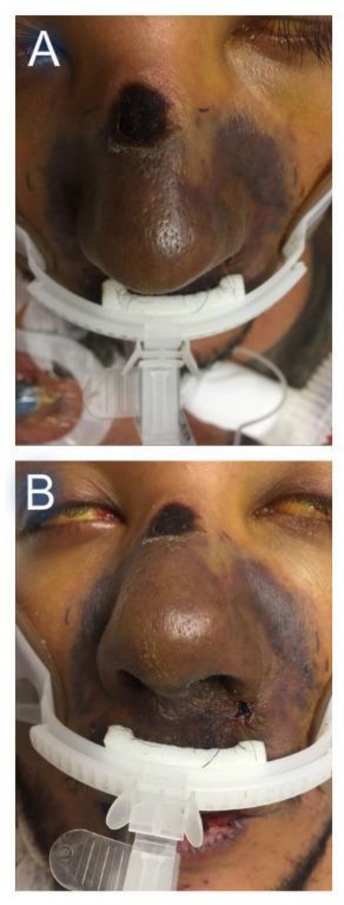
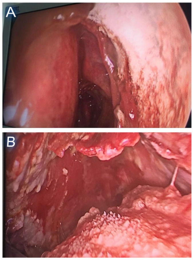
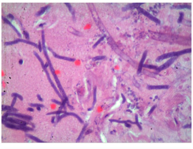
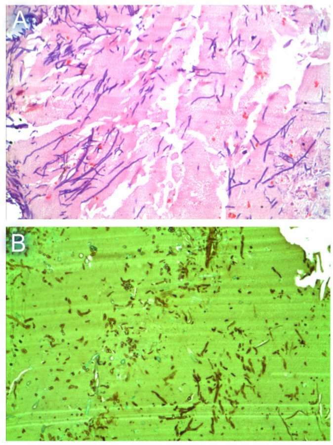
We present the case of a 28-year-old man with a long-standing history of cocaine abuse and Child-Pugh class C alcoholic liver cirrhosis who developed severe lower respiratory tract infection complicated with septic shock and multiple organ dysfunction. He was managed in the intensive care unit. On the eighth day after admission, he developed a nose discolouration, which was initially thought to be associated with high-dose vasopressors. Despite the reduction of vasopressors, the lesion progressed rapidly. It was later diagnosed as rhinocerebral mucormycosis. Amphotericin B was administered, but unfortunately the patient succumbed to the complications postinfection. The association between alcoholic liver cirrhosis and rhinocerebral mucormycosis should be known and prompt recognition warrants immediate treatment.
Keywords: adult intensive care; alcoholic liver disease; cirrhosis; infectious diseases.
© BMJ Publishing Group Ltd (unless otherwise stated in the text of the article) 2017. All rights reserved. No commercial use is permitted unless otherwise expressly granted.
Conflict of interest statement
Competing interests: None declared.
Figures
Similar articles
-
Agressive infection following a dental extraction in a diabetic patient :Rhinocerebral mucormycosis.Tunis Med. 2017 May;95(5):378-380. Tunis Med. 2017. PMID: 29509222
-
Isolated cerebral mucormycosis associated with intravenous drug use.J Mycol Med. 2020 Dec;30(4):101046. doi: 10.1016/j.mycmed.2020.101046. Epub 2020 Oct 6. J Mycol Med. 2020. PMID: 33067115
-
Cerebro-rhino orbital mucormycosis: an update.J Infect Public Health. 2012 Apr;5(2):116-26. doi: 10.1016/j.jiph.2012.01.003. Epub 2012 Mar 27. J Infect Public Health. 2012. PMID: 22541257 Review.
-
Chronic rhinocerebral mucormycosis: a rare case report and review of the literature.Mycoses. 2014 Nov;57(11):699-702. doi: 10.1111/myc.12219. Epub 2014 Jul 15. Mycoses. 2014. PMID: 25039925 Review.
-
Fatal rhinocerebral mucormycosis under the shade of hepatic encephalopathy.Ann Hepatol. 2010 Oct-Dec;9(4):462-4. Ann Hepatol. 2010. PMID: 21057167
Cited by
-
Invasive pulmonary mucormycosis and aspergillosis in a patient with decompensated hepatic cirrhosis.Med Mycol Case Rep. 2018 Mar 6;21:12-15. doi: 10.1016/j.mmcr.2018.03.004. eCollection 2018 Sep. Med Mycol Case Rep. 2018. PMID: 29560305 Free PMC article.
-
Invasive rhinocerebral mucormycosis in a patient with liver cirrhosis leading to fatal massive stroke.Med Mycol Case Rep. 2018 Sep 21;22:69-73. doi: 10.1016/j.mmcr.2018.09.006. eCollection 2018 Dec. Med Mycol Case Rep. 2018. PMID: 30294535 Free PMC article.
References
-
- Oshea R, Dasarathy S, Mccullough AJ. Alcoholic liver disease In: Practical Management of Liver Diseases. 105, 2010:98–116.
-
- Bouza E, Muñoz P, Guinea J. Mucormycosis: an emerging disease? Clin Microb Infec 2006;12:7–23. 10.1111/j.1469-0691.2006.01604.x - DOI
Publication types
MeSH terms
LinkOut - more resources
Full Text Sources
Other Literature Sources
Medical