. 2017 Oct;27(10):1195-1215.
doi: 10.1038/cr.2017.87. Epub 2017 Aug 8.

Cardiotrophin 1 stimulates beneficial myogenic and vascular remodeling of the heart

Affiliations

Cardiotrophin 1 stimulates beneficial myogenic and vascular remodeling of the heart

Mohammad Abdul-Ghani et al. Cell Res. 2017 Oct.

Abstract

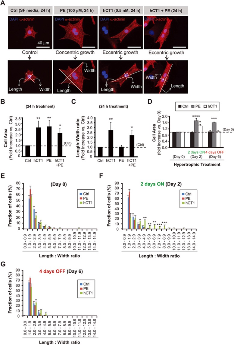
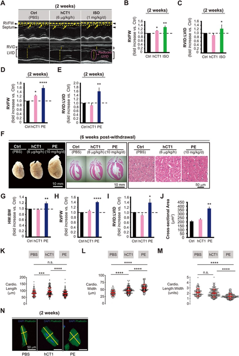
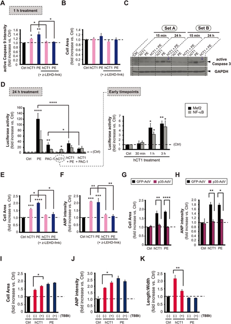
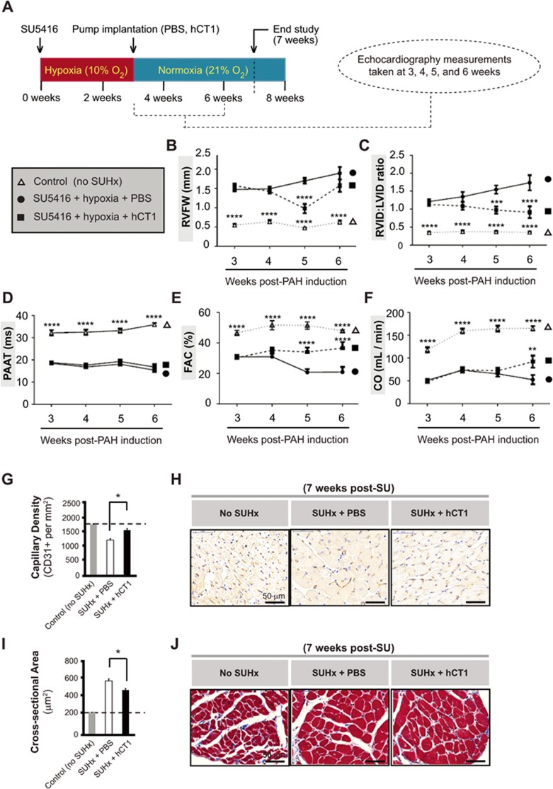
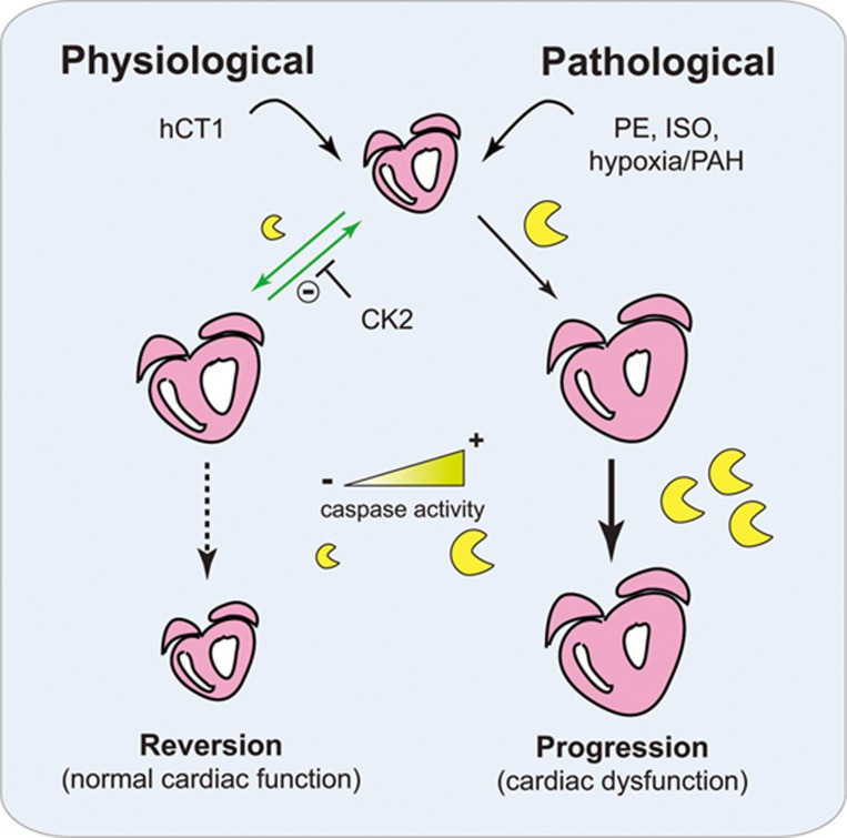
The post-natal heart adapts to stress and overload through hypertrophic growth, a process that may be pathologic or beneficial (physiologic hypertrophy). Physiologic hypertrophy improves cardiac performance in both healthy and diseased individuals, yet the mechanisms that propagate this favorable adaptation remain poorly defined. We identify the cytokine cardiotrophin 1 (CT1) as a factor capable of recapitulating the key features of physiologic growth of the heart including transient and reversible hypertrophy of the myocardium, and stimulation of cardiomyocyte-derived angiogenic signals leading to increased vascularity. The capacity of CT1 to induce physiologic hypertrophy originates from a CK2-mediated restraining of caspase activation, preventing the transition to unrestrained pathologic growth. Exogenous CT1 protein delivery attenuated pathology and restored contractile function in a severe model of right heart failure, suggesting a novel treatment option for this intractable cardiac disease.


Figures

Figure 1
Figure 1
Human cardiotrophin 1 (hCT1) induces morphologic changes in primary cardiomyocytes with reversion upon removal of hCT1 stimulation. (A) Primary neonatal cardiomyocytes were stimulated for 24 h with hCT1 (0.5 nM), PE (100 μM), hCT1 and PE, or control serum-free medium (Ctrl, SF) followed by morphometric analysis (cell area and length:width ratio). Cells were stained with α-actinin (red) and nuclei were stained with DAPI (blue). Scale bar, 40 μm. (B, C) Morphometric analysis of (A) above. Treatment with hCT1, PE, and hCT1+PE induced a significant increase in cell area versus control (n = 3; *P < 0.05 and **P < 0.01). However, only hCT1 (or hCT1+PE) stimulation resulted in elongated/eccentric growth versus control (n = 3; *P < 0.05 and **P < 0.01). (D) Cardiomyocytes were stimulated as in (A) above, but for 2 days followed by removal for 4 days. After 2 days, both hCT1 and PE induced hypertrophy versus control at day 2 (n = 3; *P < 0.05 and ****P < 0.0001). Cells reverted to pre-treatment dimensions upon removal of hCT1, whereas removal of PE caused cells to remain hypertrophied versus Control at day 6 (n = 3; ***P < 0.001). (E-G) Length:width ratio population frequency analysis of (D) above. The majority of cardiomyocytes (∼ 60%) display a length:width ratio of 1.0-2.0 (Day 0); however, upon treatment with hCT1 (Day 2), a significant shift in length:width ratio of 3.0-9.0 was observed versus control at day 2 (n = 3; **P < 0.01 and ***P < 0.001) with reversion occurring at day 6 upon hCT1 removal.
Figure 2
Figure 2
Administration of hCT1 in vivo induces beneficial cardiac growth, while withdrawal of hCT1 causes reversion to baseline cardiac dimensions. (A-J) Osmotic minipumps were implanted subcutaneously in rats containing PBS control (Ctrl), hCT1 (6 μg/kg/h), and isoproterenol (ISO, 1 mg/kg/d) or phenylephrine (PE, 10 mg/kg/d) to induce hypertrophy over 2 weeks. After 2 and 6 weeks post treatment, M-mode echocardiography analysis was used to assess myocardial structure/function. (A) Echocardiography images. Arrows and arrowheads (upper panels) point to the right ventricle free wall (RVFW) and septum. Dotted lines (lower panels) span the right and left ventricle internal diameter (RVID and LVID, respectively) of the inner chambers. (B-E) Both hCT1 and ISO (or PE) induced a significant increase in cardiac hypertrophy versus control (n = 6; *P < 0.05, **P < 0.01 and ****P < 0.0001). However, only rats treated with ISO or PE exhibited an increase in the RVID:LVID ratio (n = 6; *P < 0.05 and **P < 0.01). (F-J) 6 weeks post withdrawal, hCT1-treated rats displayed reversion of heart growth, whereas PE-treated rats maintained a significant increase in HW:BW (heart weight to body weight ratio), RVFW wall thickness, cardiomyocyte cross-sectional area, and RVID:LVID ratio versus control (n = 6; *P < 0.05, **P < 0.01 and ****P < 0.0001). (F), Whole hearts (left panel; scale bar, 10 mm), Hematoxylin and Eosin-stained sections (middle and right panels; scale bars, 10 mm and 50 μm, respectively). (K-N) Similar procedure as in (A) above, however, adult murine cardiomyocytes were analyzed. After 2 weeks of treatment, hCT1 significantly increased the length of cardiomyocytes versus PBS control or PE (K; n = 6, ***P < 0.001 and ****P < 0.0001) while PE significantly increased the width versus PBS control and hCT1 (L; n = 6, ****P < 0.0001). The length to width ratio of PE-treated cardiomyocytes was significantly decreased versus PBS control and hCT1 (M; n = 6, ****P < 0.0001), whereas no significant difference (ns) was observed between PBS control and hCT1. Representative images (N) of murine cardiomyocytes were stained with Alexa Fluor-488 Phalloidin (F-actin, green) and DAPI (nuclei, blue). Scale bar, 50 μm.
Figure 3
Figure 3
hCT1 engages a restricted activation of the intrinsic caspase-mediated cell death pathway. (A, B) Primary cardiomyocytes were treated for 1 h with control serum-free medium (Ctrl), hCT1 (0.5 nM), or PE (100 μM) in the presence or absence of the caspase 9 inhibitor (z-LEHD-fmk, 20 μM). Caspase 9 was significantly activated with hCT1 and PE versus control (n = 3; *P < 0.05 and **P < 0.01) and this was decreased in the presence of z-LEHD-fmk (n = 3; *P < 0.05). (C) Cardiomyocytes were treated with hCT1 (0.5 nM), PE (100 μM), or hCT1+PE for 15 min and 24 h and analyzed by western immunoblotting. hCT1 caused a moderate increase in caspase 3 activity compared to PE at 24 h. Caspase 3 activity also decreased with combined PE and hCT1 stimulation (arrows). GAPDH was the loading control. (D) Cardiomyocytes were transfected with reporter plasmids under the control of NF-κB or Mef2 promoters and luciferase activity was measured after treatment with: control serum-free medium (Ctrl), hCT1 (0.5 nM), PE (100 μM), or procaspase 3-activating compound 1 (PAC-1; 25 μM). hCT1 treatment resulted in NF-κB and Mef2 activation at 1 and 3 h versus control (n = 4; * P < 0.05 and **P < 0.01); and at 24 h, only NF-κB activity was sustained (n = 4; *P < 0.05). However, PE and PAC-1 treatment caused significant activation of Mef2 after 24 h (n = 4; **P < 0.01 and ****P < 0.0001). Co-stimulation of hCT1/PE and hCT1/PAC-1 significantly reduced Mef2 activity compared to PE and PAC-1 alone (n = 4; ****P < 0.0001 and *P < 0.05, respectively). (E, F) Similar procedure as in (A) above; however, with 24 h treatment. hCT1 and PE significantly increased cell area and the pro-hypertrophic marker ANP versus control (n = 3; ***P < 0.001 and ****P < 0.0001) and this was significantly attenuated in the presence of z-LEHD-fmk (n = 3;*P < 0.05 and **P < 0.01). (G, H) Cardiomyocytes were infected for 24 h with an adenovirus (AdV) encoding the caspase inhibitor, p35-AdV, prior to inducing hypertrophy with hCT1 or PE for 24 h. GFP-AdV was used as a control. p35-AdV significantly inhibited hCT1 and PE induced hypertrophy (n = 3; **P < 0.01 and ****P < 0.0001, respectively) and inhibited ANP expression (n = 3; **P < 0.01 and *P < 0.05, respectively). Both p35-AdV and GFP-AdV were used at a mean of infectivity (MOI) of 20. (I-K) Similar procedure as in (E, F) above; however, casein kinase 2 (CK2) activity was blocked using TBBt (50 μM). CK2 inhibition significantly increased cell size and ANP expression (n = 3; *P < 0.05) while reducing length:width ratio (n = 3; **P < 0.01) when compared to hCT1 treatment alone.
Figure 4
Figure 4
hCT1 enhances angiogenesis and limits fibrosis during cardiac hypertrophy. (A-D) Rats were treated with hCT1 or PE for 2 weeks followed by withdrawal for 6 weeks. Capillary density was assessed by staining for CD31 (brown) and counterstaining with Hematoxylin (blue). hCT1-treated rats displayed a significant increase in CD31-positive capillaries (n = 4;**P < 0.01) at 2 weeks with reversion at 6 weeks post withdrawal; whereas PE-treated rats displayed a significant decrease at 2 weeks post treatment and 6 weeks post withdrawal (n = 4; *P < 0.05). Scale bar, 50 μm. (E) Primary cardiomyocytes were incubated with control serum-free medium (Ctrl, SF medium), hCT1 (0.5 nM), PE (100 μM), or PAC-1 (25 μM) for 24 h. hCT1 treatment resulted in a greater increase in vascular endothelial growth factor (VEGF) expression compared to PE or PAC-1. Immunofluorescence was used to detect α-actinin (red), VEGF (green), and nuclei were stained with DAPI (blue). Scale bar, 40 μm. (F) Similar procedure as in (E) above, however, conditioned medium was harvested at 24 h and expression of secreted VEGF protein was detected upon immunoblotting with anti-VEGF antibody. A significant increase in VEGF was observed in hCT1-treated samples (sets A-C) when compared to PE or Ctrl. (+), recombinant VEGF protein (40 ng) used as positive control. (#), cross-reactivity bands of antibody to components in conditioned medium. (G-J) Rats were treated as in (A) above and fibrosis/collagen was assessed using Masson's Trichrome staining (blue deposits). Rat hearts exposed to PE exhibited a significant increase in collagen/fibrosis at 2 weeks post treatment as well as 6 weeks post withdrawal (n = 4; ***P < 0.001 and **P < 0.01, respectively). Scale bar, 50 μm.
Figure 5
Figure 5
hCT1 attenuates the morphologic and hemodynamic effects of pulmonary arterial hypertension (PAH) in the Sugen/hypoxia (SUHx) rat model. (A) PAH was induced by administering a single subcutaneous injection of SU5416 (SU) at 20 mg/kg (VEGF receptor antagonist) followed by exposure to chronic hypoxia (Hx) at 10% oxygen for 3 weeks. Rats were recovered for 24 h in normoxia before subcutaneous implantation of osmotic minipumps containing hCT1 (6 μg/kg/h) or phosphate-buffered saline (PBS). Echocardiography was conducted and the following parameters were measured: right ventricle free wall thickness (RVFW; B), right to left ventricle internal diameter ratio (RVID:LVID; C), pulmonary artery acceleration time (PAAT; D), fractional area change (FAC; E), and cardiac output (CO; F). Control rats were not exposed to SU5416 or hypoxia. (B-F) Significant differences were observed between the PAH-induced SUHx rat groups (hCT1 or PBS) and the control rat group (n = 3; ****P < 0.0001). At 5-6 weeks, hCT1-treated SUHx animals showed a significant increase in CO (F) and FAC (E), and a decrease in RVID:LVID (C) and RVFW (B) versus the PBS-treated SUHx group (n = 3; **P < 0.01, ***P < 0.001 and ****P < 0.0001 ). (G, H) hCT1 treatment significantly increased capillary density versus PBS-treated SUHx animals (n = 3; *P < 0.05). (H) Capillaries stained with endothelial-specific CD31+ antibody followed by chromogenic detection (DAB, brown). Nuclei counterstained with Hematoxylin. Scale bar, 50 μm. (I, J) hCT1 treatment significantly decreased cardiomyocyte cross-sectional area in the right ventricle versus PBS-treated SUHx animals (n = 3; *P < 0.05). (J) Masson's Trichrome staining of heart cross-sectional area. Scale bar, 50 μm.
Figure 6
Figure 6
Proposed model of hCT1-mediated physiologic cardiac hypertrophy. hCT1 promotes reversible and beneficial cardiac remodeling by restraining caspase activation via CK2; whereas pathological stimulation (with PE, ISO, or hypoxia/PAH) causes unrestricted caspase activation with progression to cardiac dysfunction. Heart (pink), caspases (yellow), hCT1 (human cardiotrophin 1), PE (phenylephrine), ISO (isoproterenol), CK2 (casein kinase 2), PAH (pulmonary arterial hypertension).

Comment in

References

    1. Hill JA, Olson EN. Cardiac plasticity. N Engl J Med 2008; 358:1370–1380. - PubMed
    1. Harvey PA, Leinwand LA. The cell biology of disease: cellular mechanisms of cardiomyopathy. J Cell Biol 2011; 194:355–365. - PMC - PubMed
    1. van Berlo JH, Maillet M, Molkentin JD. Signaling effectors underlying pathologic growth and remodeling of the heart. J Clin Invest 2013; 123:37–45. - PMC - PubMed
    1. Chung E, Leinwand LA. Pregnancy as a cardiac stress model. Cardiovasc Res 2014; 101:561–570. - PMC - PubMed
    1. Roh J, Rhee J, Chaudhari V, Rosenzweig A. The role of exercise in cardiac aging: from physiology to molecular mechanisms. Circ Res 2016; 118:279–295. - PMC - PubMed