. 2017 Aug 7;6(8):e369.
doi: 10.1038/oncsis.2017.71.

Lysine demethylase KDM2A inhibits TET2 to promote DNA methylation and silencing of tumor suppressor genes in breast cancer

Affiliations

Lysine demethylase KDM2A inhibits TET2 to promote DNA methylation and silencing of tumor suppressor genes in breast cancer

J-Y Chen et al. Oncogenesis. .

Abstract

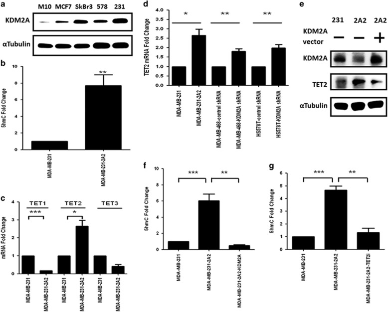
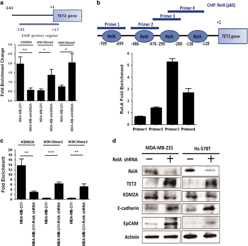
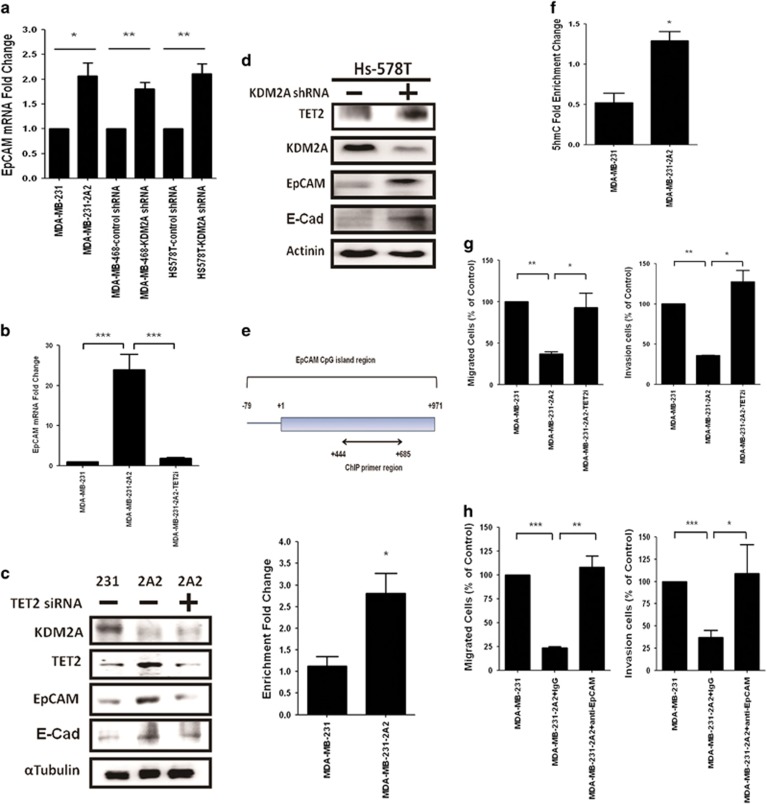
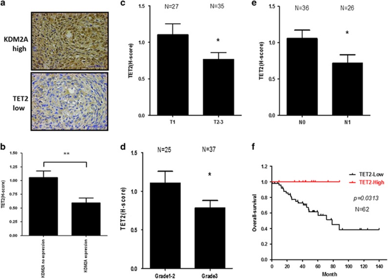
The coupling between DNA methylation and histone modification contributes to aberrant expression of oncogenes or tumor suppressor genes that leads to tumor development. Our previous study demonstrated that lysine demethylase 2A (KDM2A) functions as an oncogene in breast cancer by promoting cancer stemness and angiogenesis via activation of the Notch signaling. Here, we demonstrate that knockdown of KDM2A significantly increases the 5'-hydroxymethylcytosine (5'-hmc) level in genomic DNA and expression of tet-eleven translocation 2 (TET2) in various breast cancer cell lines. Conversely, ectopic expression of KDM2A inhibits TET2 expression in KDM2A-depleted cells suggesting TET2 is a transcriptional repression target of KDM2A. Our results show that KDM2A interacts with RelA to co-occupy at the TET2 gene promoter to repress transcription and depletion of RelA or KDM2A restores TET2 expression. Upregulation of TET2 in the KDM2A-depleted cells induces the re-activation of two TET downstream tumor suppressor genes, epithelial cell adhesion molecule (EpCAM) and E-cadherin, and inhibits migration and invasion. On the contrary, knockdown of TET2 in these cells decreases EpCAM and E-cadherin and increases cell invasiveness. More importantly, TET2 expression is negatively associated KDM2A in triple-negative breast tumor tissues, and its expression predicts a better survival. Taken together, we demonstrate for the first time that TET2 is a direct repression target of KDM2A and reveal a novel mechanism by which KDM2A promotes DNA methylation and breast cancer progression via the inhibition of a DNA demethylase.


Conflict of interest statement

The authors declare no conflict of interest.

Figures

Figure 1
Figure 1
KDM2A inhibits the expression of TET2 to reduce the 5′-hmc level in breast cancer cells. (a) Cellular proteins were extracted from various breast cancer cell lines with a lysis buffer (50 mM Tris-HCl, pH 7.4, 150 mM NaCl, 1% NP-40, 0.1% sodium dodecyl sulfate (SDS), 0.5% sodium deoxycholate, 2 mM ethylenediaminetetraacetic acid (EDTA) and 50 mM sodium fluoride (NaF)) and the proteins were separated by SDS-polyacrylamide gel electrophoresis. Proteins were transferred to polyvinylidene difluoride (PVDF) membranes, probed with KDM2A antibody (Abcam, Cambridge, MA, USA) and the signal was developed by enhanced chemiluminescence reagent. α-Tubulin was used as an internal control. (b) Genomic DNA of MDA-MB-231 and KDM2A-depleted MDA-MB-231-2A2 cells was extracted by the Tissue & Cell Genomic DNA purification kit (GMbiolab Co. Ltd, Taiwan). The 5′-hydroxymethylcytosine (5′-hmc) level of genomic DNA was detected by using Quest 5-hmC TM DNA ELISA Kit (ZYMO Research Corp. Irvine, USA). Results from three independent assays were collected and the 5’-hmc level of MD-MB-231 cells was defined as 1. (c) Total RNA was isolated from cells, and 1 μg of RNA was reverse-transcripted to cDNA. Target mRNAs were quantified using real-time PCR reactions with SYBR green fluorescein and actin was served as an internal control. Primer sequences used for real-time PCR was showed in Supplementary Table 1. Data were shown as Mean±s.e.m. (d) MDA-MB-468 and Hs758T breast cancer cells were transfected with KDM2A shRNA and the mRNA level of TET2 was determined at 48 h after transfection. (e) MDA-MB-231 (231) or MDA-MB-231-2A2 (2A2) cells were transfected with control (—) or KDM2A expression vector. After 48 h, protein level of KDM2A and TET2 was studied by western blotting. (f) MDA-MB-231-2A2 cells were transfected with KDM2A expression vector and the 5′-hmc level of genomic DNA was determined by using Quest 5-hmC TM DNA ELISA Kit. (g) MDA-MB-231 or MDA-MB-231-2A2 cells were transfected without or with TET2 siRNA (Santa Cruz Biotechnology, Inc., USA), and the 5′-hmc level of genomic DNA was determined by using Quest 5-hmC TM DNA ELISA Kit. Statistical analysis was performed by using paired t-test and two-tailed P-values ⩽0.05 were considered statistically significant. ***P<0.001, **P<0.01, *P<0.05.
Figure 2
Figure 2
RelA is involved in the inhibition of TET2 by KDM2A. (a) The diagram in the upper panel showed the genomic region of human TET2 gene amplified in our ChIP assay. MDA-MB-231 cells were transfected with control (—) or KDM2A shRNA. After 48 h, cells were fixed with 1% formaldehyde at 37 °C for 10 min and washed twice with ice-cold PBS containing protease inhibitors. Cells were incubated in a lysis buffer (1% SDS, 10 mM EDTA, 50 mM Tris-HCl, pH 8.1) for 10 min on ice and sonicated to shear genomic DNA. The lysate was centrifuged for 10 min at 13 000 r.p.m. at 4 °C. The supernatant was diluted in a ChIP dilution buffer (0.01% SDS, 1% Triton X-100, 2 mM EDTA, 16.7 mM Tris-HCl, pH 8.1, 167 mM NaCl, and protease inhibitors). Anti-KDM2A, anti-dimethyl H3K36, anti-trimethyl H3K36 and non-immune (negative control) antibodies were added to the supernatant and incubated overnight at 4 °C with rotation. DNA fragments were recovered and subjected to PCR amplification. List of primer sequences used for ChIP assay was showed in Supplementary Table 1. (b) Transfection factor binding sites in the human TET2 gene promoter region were predicted by PROMO software (http://alggen.lsi.upc.es/) and the four potential RelA binding sites were shown in the upper panel. ChIP assay was carried out as described in (a) by using anti-RelA antibody (Thermo Fisher Scientific Inc., Waltham, MA, USA). The relative enrichment of RelA binding to the four potential sites was shown. (c) MDA-MB-231 cells were transfected with control or RelA shRNA. After 48 h, ChIP assay was conducted to investigate the binding of KDM2A to proximal TET2 gene promoter shown in (a). The methylation status (demethylation and trimethylation) of H3K36 in this region was also studied by ChIP assay. (d) MDA-MB-231 and Hs-578T cells were transfected with control (—) or RelA shRNA. The protein level of RelA, TET2 and KDM2A was studied by western blotting at 48 h after transfection.
Figure 3
Figure 3
Increase of TET2 induced by KDM2A depletion re-activates the downstream target genes and attenuates cell invasiveness. (a) MDA-MB-231, MDA-MB-468 and Hs-578T cells were transfected with control (—) or KDM2A shRNA. After 48 h, the expression of EpCAM was studied by real-time RT–PCR and was compared between control and KDM2A-depleted cells. (b) MDA-MB-231-2A2 cells were transfected without or with TET2 siRNA and the expression of EpCAM was investigated by real-time RT–PCR at 48 h after transfection. (c) Protein level of two reported TET target genes EpCAM and E-cadherin in MDA-MB-231 cells and MDA-MB-231-2A2 cells transfected without or with TET2 siRNA was also studied by western blotting. (d) Hs-578T cells were transfected with control (—) or KDM2A shRNA. After 48 h, protein level of EpCAM and E-cadherin was investigated. (e) The upper diagram showed the prediction of a CpG island (−79 to +971) in human EpCAM promoter by the University of California Santa Cruz genome browser (https://genome.ucsc.edu) and the region (+444 to +685) amplified by our PCR primer was shown. The binding of TET2 to this CpG region in MDA-MB-231 and MDA-MB-231-2A2 cells was studied by ChIP assay. (f) The 5′-hmc level in the amplified region was also investigated by using anti-5′-hmc antibody for ChIP assay. (g) MDA-MB-231 or MDA-MB-231-2A2 cells were transfected without or with TET2 siRNA. Migration assays were carried out in transwells with 5-μm pore filter inserts on 24-well plates. For invasion assays, the transwell inserts were coated with gelatin A/B solution before the cells were seeded. The lower chamber was filled with medium containing 1% serum. After 12 h, the filter was gently removed from the chamber, the cells on the upper surface were removed by wiping with a cotton swab, and cells that migrated to the lower surface areas were fixed, stained with DAPI and counted in 15 randomly selected fields in a microscope. Experiments were repeated three times. (h) MDA-MB-231 and MDA-MB-231-2A2 cells were incubated with non-immune IgG or anti-EpCAM antibody and subjected to migration and invasion assays as described in (f). **P<0.01, *P<0.05.
Figure 4
Figure 4
TET2 expression is negatively associated with KDM2A and predicts a better survival in triple-negative breast cancer patients. (a) Typical immunohistochemical staining showed strong KDM2A and low TET2 expression in a tumor tissue. Paraffin-embedded tissue sections of 62 human triple-negative breast cancer specimens were obtained from Department of Pathology, Kaohsiung Medical University Hospital (Kaohsiung, Taiwan). The slides were stained with anti-KDM2A and anti-TET2 antibody and the staining was interpreted using the H-score, defined by the following equation: H-score=ΣPi (i + 1) as previously described. Institutional review board approval for using these human tissues in this study was given by the Research Ethics Committee of the Kaohsiung Medical Hospital (IRB: KMUHIRB-E(II)-20150086). (b) The association between the expression of KDM2A and TET2 was compared. In addition, the expression of TET2 in patients with different tumor sizes (c), grade (d) and lymph node metastasis (e) was compared. (f) Patient’s overall survival was compared by the Kaplan–Meier plots and compared using the log-rank test. **P<0.01, *P<0.05.

References

    1. Cedar H, Bergman Y. Linking DNA methylation and histone modification: patterns and paradigms. Nat Rev Genet 2009; 10: 295–304. - PubMed
    1. Chi P, Allis CD, Wang GG. Covalent histone modifications—miswritten, misinterpreted and mis-erased in human cancers. Nat Rev Cancer 2010; 10: 457–469. - PMC - PubMed
    1. Brien GL, Valerio DG, Armstrong SA. Exploiting the epigenome to control cancer-promoting gene-expression programs. Cancer Cell 2016; 29: 464–476. - PMC - PubMed
    1. Ito S, D'Alessio AC, Taranova OV, Hong K, Sowers LC, Zhang Y. Role of Tet proteins in 5mC to 5hmC conversion, ES-cell self-renewal and inner cell mass specification. Nature 2010; 466: 1129–1133. - PMC - PubMed
    1. Ito S, Shen L, Dai Q, Wu SC, Collins LB, Swenberg JA et al. Tet proteins can convert 5-methylcytosine to 5-formylcytosine and 5-carboxylcytosine. Science 2011; 333: 1300–1303. - PMC - PubMed

LinkOut - more resources