

Very Rare Amelanotic Lentigo Maligna Melanoma with Skull Roof Invasion
- PMID: 28785332
- PMCID: PMC5535657
- DOI: 10.3889/oamjms.2017.113
Very Rare Amelanotic Lentigo Maligna Melanoma with Skull Roof Invasion
Abstract
Background: Lentigo malignant melanoma is a melanoma subtype of chronic sun-damaged skin in elderly Caucasians. Amelanotic variants of lentigo malignant are extremely rare.
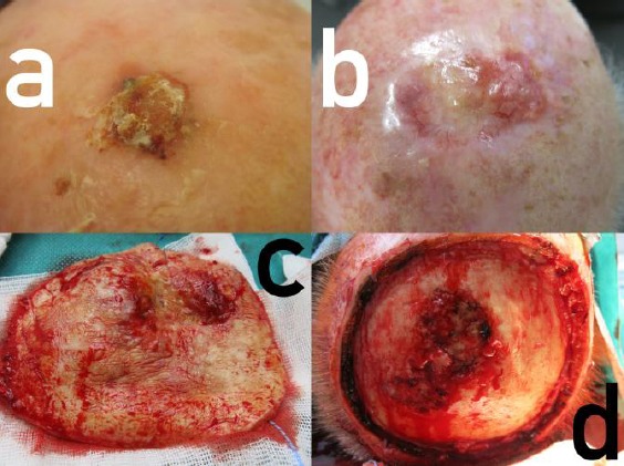
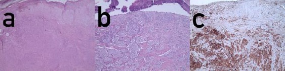
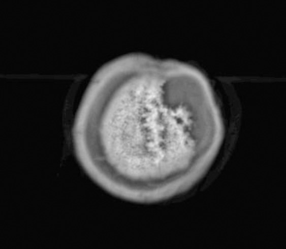
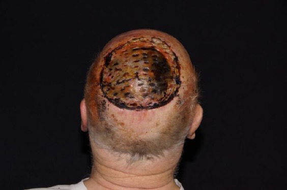
Case presentation: This is a case report of an 80-year-old male patient who presented with a non-pigmented exophytic tumour of his bald head. After complete surgical excision under the suspicion of squamous cell carcinoma, three-dimensional histologic examination confirmed an amelanotic lentigo malignant melanoma with a tumour thickness of 1.76 mm, resected R0. Five years later he developed the first relapse, the other year a satellite metastasis was surgically removed. One year later, this patient had developed a large relapsing lentigo malignant melanoma with skull roof invasion. There was no evidence of distant metastatic spread. Amelanotic lentigo malignant melanoma is a very rare tumour.
Conclusions: Serial excision or slow Mohs and Mohs micrographic surgery are the treatments of choice especially in the head and neck area. These tumours may be locally very aggressive as it is shown by skull invasion in the present case.
Keywords: Melanoma; amelanotic lentigo malignant melanoma; follow-up; relapses; skull invasion; surgery.
Figures
Similar articles
-
Amelanotic Lentigo Maligna Melanoma: Mohs Surgery as the Definitive Treatment of an Invisible Tumour.J Cutan Med Surg. 2018 Jan/Feb;22(1):51-57. doi: 10.1177/1203475417719046. Epub 2017 Jul 7. J Cutan Med Surg. 2018. PMID: 28685596
-
Recurrent lentigo maligna as amelanotic lentigo maligna melanoma.J Eur Acad Dermatol Venereol. 2002 Sep;16(5):506-10. doi: 10.1046/j.1468-3083.2002.00534.x. J Eur Acad Dermatol Venereol. 2002. PMID: 12428849
-
Role of In Vivo Reflectance Confocal Microscopy in the Analysis of Melanocytic Lesions.Acta Dermatovenerol Croat. 2018 Apr;26(1):64-67. Acta Dermatovenerol Croat. 2018. PMID: 29782304 Review.
-
Amelanotic lentigo maligna melanoma: a unique case presentation.Dermatol Surg. 1999 May;25(5):408-11. doi: 10.1046/j.1524-4725.1999.08271.x. Dermatol Surg. 1999. PMID: 10469082
-
Amelanotic lentigo maligna melanoma: a unique case and review of the literature.Cutis. 1989 Jul;44(1):45-8. Cutis. 1989. PMID: 2666033 Review.
Cited by
-
Nonmelanoma Skin Cancer with Skull Infiltration and Cranial Involvement.Open Access Maced J Med Sci. 2019 Sep 30;7(18):3030-3033. doi: 10.3889/oamjms.2019.416. eCollection 2019 Sep 30. Open Access Maced J Med Sci. 2019. PMID: 31850116 Free PMC article.
References
-
- Juhász ML, Marmur ES. Reviewing challenges in the diagnosis and treatment of lentigo maligna and lentigo-maligna melanoma. Rare Cancers Ther. 2015;3:133–145. https://doi.org/10.1007/s40487-015-0012-9 PMid:27182482 PMCid:PMC4837936. - PMC - PubMed
-
- Kvaskoff M, Siskind V, Green AC. Risk factors for lentigo maligna melanoma compared with superficial spreading melanoma:a case-control study in Australia. Arch Dermatol. 2012;148(2):164–170. https://doi.org/10.1001/archdermatol.2011.291 PMid:22004881. - PubMed
-
- Greveling K, Wakkee M, Nijsten T, van den Bos RR, Hollestein LM. Epidemiology of lentigo maligna and lentigo maligna melanoma in the Netherlands 1989-2013. J Invest Dermatol. 2016;136(10):1955–1960. https://doi.org/10.1016/j.jid.2016.06.014 PMid:27349862. - PubMed
-
- Christensen KN, Hochwalt PC, Hocker TL, Roenigk RK, Brewer JD, Baum CL, Otley CC, Arpey CJ. Comparison of MITF and Melan-A immunohistochemistry during Mohs surgery for lentigo maligna-type melanoma in situ and lentigo maligna melanoma. Dermatol Surg. 2016;42(2):167–175. https://doi.org/10.1097/DSS.0000000000000600 PMid:26771682. - PubMed
-
- Jaimes N, Marghoob AA, Rabinovitz H, Braun RP, Cameron A, Rosendahl C, Canning G, Keir J. Clinical and dermoscopic characteristics of melanomas on nonfacial chronically sun-damaged skin. J Am Acad Dermatol. 2015;72(6):1027–1035. https://doi.org/10.1016/j.jaad.2015.02.1117 PMid:25824275. - PubMed
Publication types
LinkOut - more resources
Full Text Sources
Other Literature Sources