Case Reports
. 2017:2017:2897617.
doi: 10.1155/2017/2897617. Epub 2017 Jul 12.

Different Clinical Features of Acral Abortive Hemangiomas

Affiliations
Case Reports

Different Clinical Features of Acral Abortive Hemangiomas

N Vega Mata et al. Case Rep Dermatol Med. 2017.

Abstract

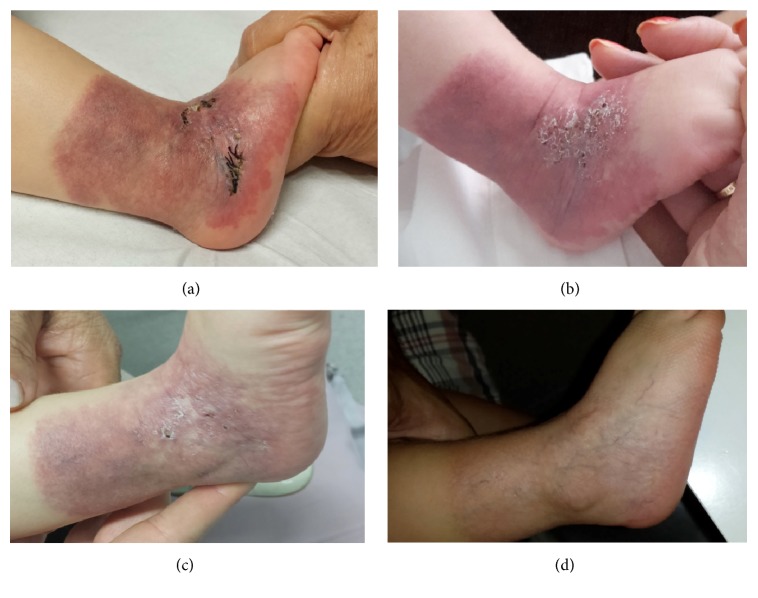
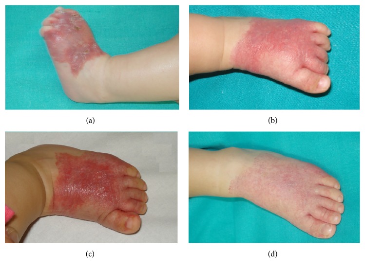
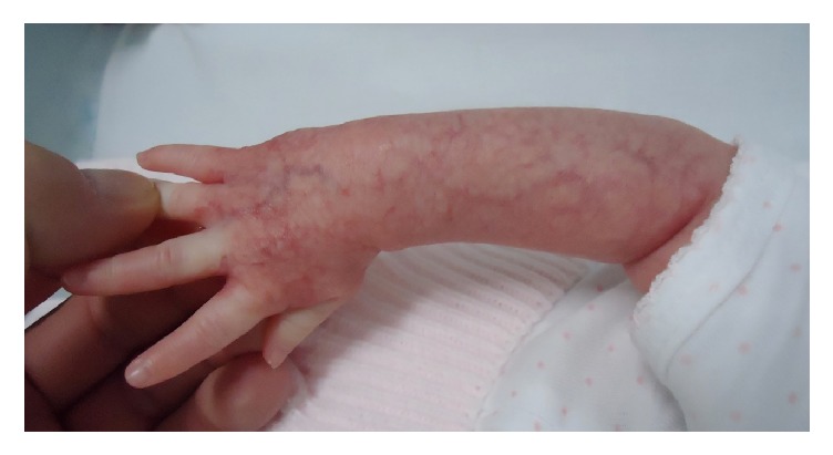
Some infantile hemangiomas called in literature "minimal or arrested growth hemangiomas" or "abortive hemangiomas" are present at birth and have a proliferative component equaling less than 25% of its total surface area. Often, they are mistaken for vascular malformation. We present five patients (three girls and two boys) with abortive hemangiomas diagnosed between January 2010 and December 2015 localized in acral part of the extremities. They were congenital lesions resembling precursor of hemangiomas but did not show proliferation phase. Immunohistochemical Glut-1 was performed in all of them as a way to confirm the abortive hemangioma diagnosis. The most common appearance was a reticulated erythematous patch with multiple fine telangiectasias on the surface. We remark that one of them presented a segmental patch with two different morphologies and evolutions. The proximal part showed pebbled patches of bright-red hemangioma and presented proliferation and the distal part with a reticulated network-like telangiectasia morphology remained unchanged. We detected lower half of the body preference and dorsal region involvement preference without ventral involvement. The ulceration occurred in three patients with two different degrees of severity.


Figures

Figure 1
Figure 1
Case  1. (a) Ulceration on an erythematous patch with a network-like and blotchy appearance in the dorsal surface of the foot of a boy of 3 months. (b) Resolution of the ulceration after 1 month with propranolol treatment. Progressive regression of the lesion at 9 months (c) and 11 months (d).
Figure 2
Figure 2
Case  2. (a) A skin biopsy on erythematous scaly plaque with geometric border covering all lateral aspect of her right ankle as a sock-like distribution. (b) Numerous superficial ulcerations of this erythematous scaly plaque. (c) Progressive regression of the lesion and the ulceration with propranolol treatment. (d) Spontaneous involution of the lesion.
Figure 3
Figure 3
Case  3. (a) An erythematous scaly patch in the dorsal and lateral surface of the left foot extending onto the digits which presented multiple and superficial ulcerations. (b) Response to propranolol after the first month of treatment. (c) Response to propranolol at sixth months of treatment. (d) Spontaneous involution.
Figure 4
Figure 4
Case  4. Pebbled patches of bright-red hemangioma and the distal part extending onto the digits presenting reticulated network-like telangiectasia morphology.
Figure 5
Figure 5
Case  5. An erythematous patch with prominent surface telangiectases on the dorsal arm of a 3-month girl.

References

    1. Corella F., Garcia-Navarro X., Ribe A., Alomar A., Baselga E. Abortive or minimal-growth hemangiomas: Immunohistochemical evidence that they represent true infantile hemangiomas. Journal of the American Academy of Dermatology. 2008;58(4):685–690. doi: 10.1016/j.jaad.2007.08.007. - DOI - PubMed
    1. Toledo-Alberola F., Betlloch-Mas I., Cuesta-Montero L., et al. Abortive hemangiomas. Description of clinical and pathological findings with special emphasis on dermoscopy. European Journal of Dermatology. 2010;20(4):497–500. doi: 10.1684/ejd.2010.0959. - DOI - PubMed
    1. North P. E., Waner M., Mizeracki A., Mihm M. C., Jr. GLUT1: A newly discovered immunohistochemical marker for juvenile hemangiomas. Human Pathology. 2000;31(1):11–22. doi: 10.1016/S0046-8177(00)80192-6. - DOI - PubMed
    1. Martín-Santiago A., Bauzá A., del Pozo L., Carrillo P. Abortive or Minimal-Growth Hemangiomas. A Review of 14 Cases. Actas Dermosifiliogr. 2012;103(3):246–250. doi: 10.1016/j.adengl.2012.04.007. - DOI - PubMed
    1. Liang M. G., Frieden I. J. Infantile and congenital hemangiomas. Seminars in Pediatric Surgery. 2014;23(4):162–167. doi: 10.1053/j.sempedsurg.2014.06.017. - DOI - PubMed

Publication types

LinkOut - more resources