

Measurement of the blood flow rate and velocity in coronary artery stenosis using intracoronary frequency domain optical coherence tomography: Validation against fractional flow reserve
- PMID: 28785616
- PMCID: PMC5497166
- DOI: 10.1016/j.ijcha.2014.10.004
Measurement of the blood flow rate and velocity in coronary artery stenosis using intracoronary frequency domain optical coherence tomography: Validation against fractional flow reserve
Abstract
Objectives: The main objective of this study was to assess the blood flow rate and velocity in coronary artery stenosis using intracoronary frequency domain optical coherence tomography (FD-OCT). A correlation between fractional flow reserve (FFR) and FD-OCT derived blood flow velocity is also included in this study.
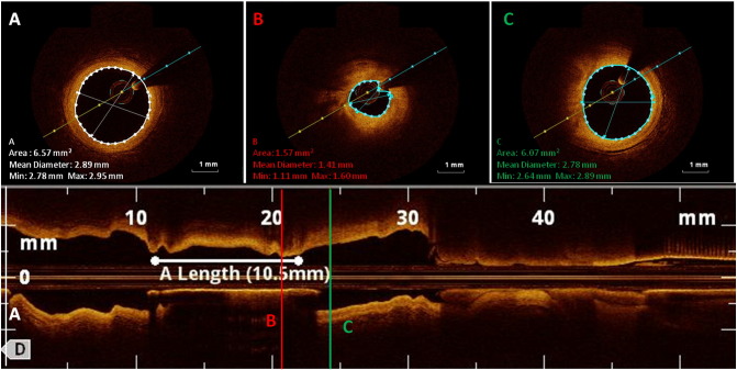
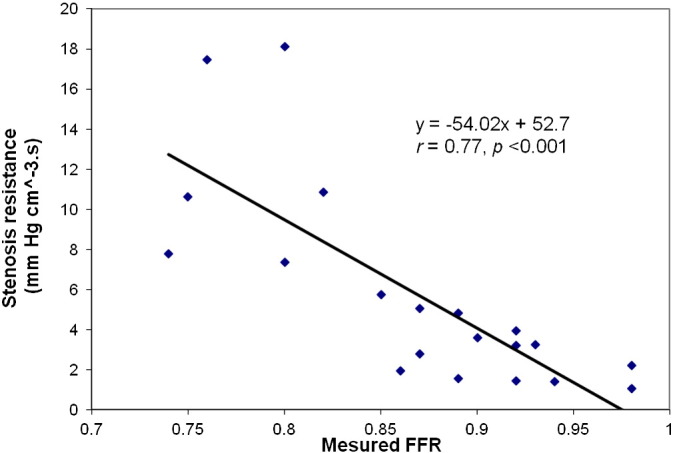
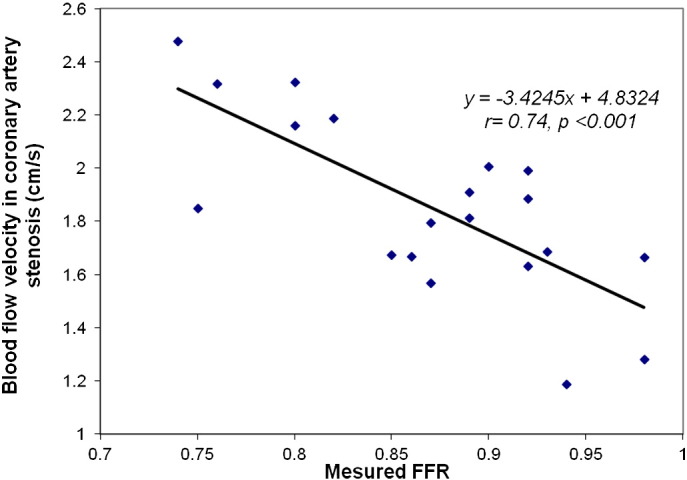
Methods & results: A total of 20 coronary stenoses in 15 patients were assessed consecutively by quantitative coronary angiography (QCA), FFR and FD-OCT. A percutaneous coronary intervention (PCI) optimization system was used in this study which combines wireless FFR measurement and FD-OCT imaging in one platform. Stenoses were labelled severe if FFR ≤ 0.8. Blood flow rate and velocity in each stenosis segment were derived from the volumetric analysis of the FD-OCT pull back images. The FFR value was ≤ 0.80 in 5 stenoses (25%). The mean blood flow rate in severe coronary stenosis (n = 5) was 2.54 ± 0.55 ml/s as compared to 4.81 ± 1.95 ml/s in stenosis with FFR > 0.8 (n = 15). A good and significant correlation between FFR and FD-OCT blood flow velocity in coronary artery stenosis (r = 0.74, p < 0.001) was found.
Conclusion: The assessment of stenosis severity using FD-OCT derived blood flow rate and velocity has the ability to overcome many limitations of QCA and intravascular ultrasound (IVUS).
Keywords: Blood flow rate; Blood flow velocity; Coronary lesions; Fractional flow reserve (FFR); Intracoronary optical coherence tomography (IOCT).
Figures
References
-
- Pijls N.H., Fearon W.F., Tonino P.A., Siebert U., Ikeno F., Bornschein B. Fractional flow reserve versus angiography for guiding percutaneous coronary intervention in patients with multivessel coronary artery disease: 2-year follow-up of the FAME (Fractional Flow Reserve Versus Angiography for Multivessel Evaluation) study. J Am Coll Cardiol. 2010;56(3):177–184. - PubMed
-
- Tonino P.A., De Bruyne B., Pijls N.H., Siebert U., Ikeno F., van' t Veer M. Fractional flow reserve versus angiography for guiding percutaneous coronary intervention. N Engl J Med. 2009;360(3):213–224. - PubMed
-
- White C.W., Wright C.B., Doty D.B., Hiratza L.F., Eastham C.L., Harrison D.G. Does visual interpretation of the coronary arteriogram predict the physiologic importance of a coronary stenosis? N Engl J Med. 1984;310(13):819–824. - PubMed
-
- De Bruyne B., Pijls N.H., Bartunek J., Kulecki K., Bech J.W., De Winter H. Fractional flow reserve in patients with prior myocardial infarction. Circulation. 2001;104(2):157–162. - PubMed
-
- Pijls N.H., Van Gelder B., Van der Voort P., Peels K., Bracke F.A., Bonnier H.J. Fractional flow reserve: a useful index to evaluate the influence of an epicardial coronary stenosis on myocardial blood flow. Circulation. 1995;92(11):3183–3193. - PubMed
LinkOut - more resources
Full Text Sources
Other Literature Sources
Miscellaneous
