

Cardiac calcified amorphous tumor: A systematic review of the literature
- PMID: 28785635
- PMCID: PMC5497183
- DOI: 10.1016/j.ijcha.2015.01.012
Cardiac calcified amorphous tumor: A systematic review of the literature
Abstract
Background: Calcified amorphous tumor (CAT) of the heart is a rare non-neoplastic intracavitary cardiac mass. Several case reports have been published but large series are lacking.
Objective: To determine clinical features, current management and outcomes of this rare disease.
Design: A systematic review of all articles reporting cases of CAT in order to perform a pooled analysis of its clinical features, management and outcomes.
Data sources: An electronic search of all English articles using PUBMED was performed. Further studies were identified by cross-referencing from relevant papers.
Inclusion criteria: We restricted inclusion to articles reporting cases of CAT in the English language literature published up to July 2014.
Data extraction: One author performed data extraction using predefined data fields.
Results: A total of 27 articles, reporting 42 cases of CAT were found and included in this review.
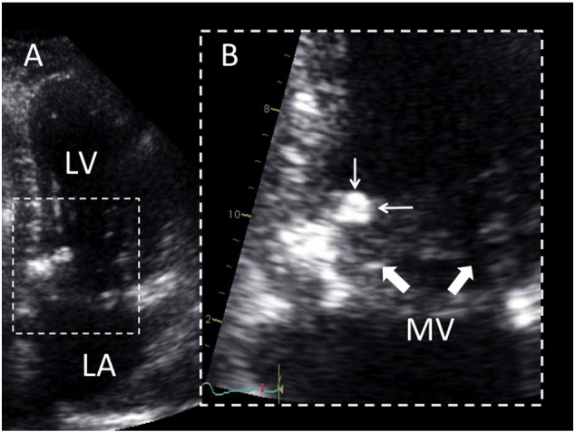
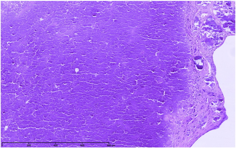
Conclusion: In this review, the most frequent presenting symptoms were dyspnea and embolic events. Mitral valve and annulus were the most frequent location of CAT. Surgery was most of the time required to confirm diagnosis, and was relatively safe. Overall outcome after surgical resection was good.
Keywords: Calcified amorphous tumor; Heart; Mass; Nonneoplastic; Review.
Figures
References
-
- Reynolds C., Tazelaar H.D., Edwards W.D. Calcified amorphous tumor of the heart (cardiac CAT) Hum Pathol. 1997;28:601–606. - PubMed
-
- Vlasseros I., Katsi V., Tousoulis D., Tsiachris D., Bousiotou A., Souretis G. Visual loss due to cardiac calcified amorphous tumor: a case report and brief review of the literature. Int J Cardiol. 2011;152:e56–e57. - PubMed
-
- Ho H.H., Min J.K., Lin F., Wong S.C., Bergman G. Images in cardiovascular medicine. Calcified amorphous tumor of the heart. Circulation. 2008;117:e171–e172. - PubMed
-
- Habib A., Friedman P.A., Cooper L.T., Suleiman M., Asirvatham S.J. Cardiac calcified amorphous tumor in a patient presenting for ventricular tachycardia ablation: intracardiac echocardiogram diagnosis and management. J Interv Card Electrophysiol. 2010;29:175–178. - PubMed
LinkOut - more resources
Full Text Sources
Other Literature Sources
Miscellaneous
