

Residual Deep Convolutional Neural Network Predicts MGMT Methylation Status
- PMID: 28785873
- PMCID: PMC5603430
- DOI: 10.1007/s10278-017-0009-z
Residual Deep Convolutional Neural Network Predicts MGMT Methylation Status
Abstract
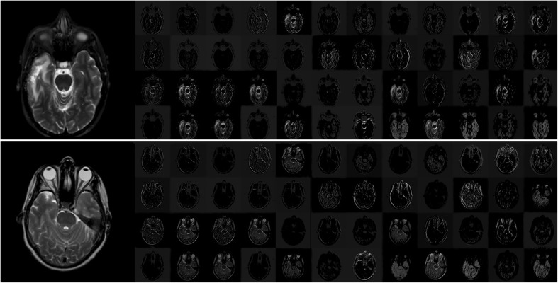
Predicting methylation of the O6-methylguanine methyltransferase (MGMT) gene status utilizing MRI imaging is of high importance since it is a predictor of response and prognosis in brain tumors. In this study, we compare three different residual deep neural network (ResNet) architectures to evaluate their ability in predicting MGMT methylation status without the need for a distinct tumor segmentation step. We found that the ResNet50 (50 layers) architecture was the best performing model, achieving an accuracy of 94.90% (+/- 3.92%) for the test set (classification of a slice as no tumor, methylated MGMT, or non-methylated). ResNet34 (34 layers) achieved 80.72% (+/- 13.61%) while ResNet18 (18 layers) accuracy was 76.75% (+/- 20.67%). ResNet50 performance was statistically significantly better than both ResNet18 and ResNet34 architectures (p < 0.001). We report a method that alleviates the need of extensive preprocessing and acts as a proof of concept that deep neural architectures can be used to predict molecular biomarkers from routine medical images.
Keywords: Deep learning; MGMT methylation; MRI.
Figures

References
-
- Weizman L, Ben-Sira L, Joskowicz L, Aizenstein O, Shofty B, Constantini S, Ben-Bashat D. Prediction of brain MR scans in longitudinal tumor follow-up studies. Med Image Comput Comput Assist Interv. 2012;15:179–187. - PubMed
-
- Law M, Young RJ, Babb JS, Peccerelli N, Chheang S, Gruber ML, Miller DC, Golfinos JG, Zagzag D, Johnson G. Gliomas: predicting time to progression or survival with cerebral blood volume measurements at dynamic susceptibility-weighted contrast-enhanced perfusion MR imaging. Radiology. 2008;247:490–498. doi: 10.1148/radiol.2472070898. - DOI - PMC - PubMed
MeSH terms
Substances
Grants and funding
LinkOut - more resources
Full Text Sources
Other Literature Sources
Medical
Research Materials
