Review
. 2017 Oct;8(5):483-489.
doi: 10.1007/s13244-017-0565-2. Epub 2017 Aug 7.

Pulmonary fibrosis on the lateral chest radiograph: Kerley D lines revisited

Affiliations
Review

Pulmonary fibrosis on the lateral chest radiograph: Kerley D lines revisited

Daniel B Green et al. Insights Imaging. 2017 Oct.

Abstract

The retrosternal clear space (RCS) is a lucent area on the lateral chest radiograph located directly behind the sternum. The two types of pathology classically addressed in the RCS are anterior mediastinal masses and emphysema. Diseases of the pulmonary interstitium are a third type of pathology that can be seen in the RCS. Retrosternal reticular opacities, known as Kerley D lines, were initially described in the setting of interstitial oedema. Pulmonary fibrosis is another aetiology of Kerley D lines, which may be more easily identified in the RCS than elsewhere on the chest radiograph.

Teaching points: • The RCS is one of three lucent spaces on the lateral chest radiograph. • Reticular opacities in the RCS are known as Kerley D lines. • Pulmonary fibrosis can be seen in the RCS as Kerley D lines. • Kerley D lines should be further evaluated with chest CT.

Keywords: Lung diseases, interstitial; Multidetector computed tomography; Pulmonary emphysema; Pulmonary fibrosis; Thoracic radiography.


Figures

Fig. 1
Fig. 1
Lateral chest radiographs demonstrating a normal retrosternal clear space (a), the lucent area between the sternum and ascending aorta, and retrosternal reticular opacities due to interstitial pulmonary oedema, known as “Kerley D lines” (b). Pleural effusions accompany the oedema
Fig. 2
Fig. 2
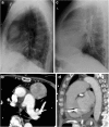
Two examples of classic retrosternal clear space pathology: a lateral chest radiograph (a) and axial CT (b) showing an anterior mediastinal mass, and a lateral chest radiograph (c) and sagittal CT (d) showing an enlarged right ventricle and main pulmonary artery in a patient with pulmonary hypertension
Fig. 3
Fig. 3
53-year-old woman with chronic hypersensitivity pneumonitis. The lateral chest radiograph (a) projects fibrosis as well through the retrosternal clear space as at the lung bases. A corresponding sagittal CT slice (b) is also shown
Fig. 4
Fig. 4
68-year-old man with idiopathic pulmonary fibrosis. An axial CT slice (a) shows fibrosis at the lung bases. On the lateral radiograph (b), reticular opacities are seen in the retrosternal clear space but not at the lung bases, where they are obscured by pulmonary vasculature and the spine. A corresponding sagittal CT slice (c) is also shown
Fig. 5
Fig. 5
63-year-old asymptomatic man with a normal radiograph (a) showing fine linear opacities in the retrosternal clear space that taper before reaching the sternum. In contrast, coarse reticular opacities form a mesh and extend to the sternum (b) in a 67-year-old man with an unclassified pulmonary fibrosis (CT not shown)
Fig. 6
Fig. 6
67-year-old man with mild pulmonary fibrosis anteriorly and posteriorly on a sagittal CT slice (a). On the lateral radiograph (b), fibrosis is only evident anteriorly, despite the small size of the retrosternal clear space. The frontal radiograph (c) is normal
Fig. 7
Fig. 7
84-year-old woman with idiopathic pulmonary fibrosis. On the lateral radiograph (a), reticular opacities are easily seen through an opaque retrosternal space. Although the retrocardiac space is more lucent, reticular opacities are not seen at the lung bases, despite the basilar predominance typical of UIP demonstrated on axial (b) and sagittal (c) CT slices
Fig. 8
Fig. 8
62-year-old woman with metastatic lung adenocarcinoma and lymphangitic carcinomatosis. Interlobular septal thickening is clearly seen in the retrosternal clear space on the lateral radiograph (a). A corresponding sagittal CT slice (b) is also shown
Fig. 9
Fig. 9
55-year-old man with smoking history. The frontal radiograph (a) is normal, and the lateral radiograph (b) shows reticular opacities in the retrosternal clear space only. Axial (c) and sagittal (d) CT slices show paraseptal and centrilobular emphysema corresponding to the radiographic abnormality. Ground-glass opacities in the lower lobes are suspected to be due to desquamative interstitial pneumonia

Similar articles

References

    1. Proto AV, Speckman JM. The left lateral radiograph of the chest. Med Radiogr Photogr. 1980;56(3):38–64. - PubMed
    1. Heitzman ER, Markarian B, Solomon J. Chronic obstructive pulmonary disease. A review, emphasizing roentgen pathologic correlations. Radiol Clin N Am. 1973;11(1):49–75. - PubMed
    1. Webb WR, Higgins CB. (2010) Thoracic Imaging: Pulmonary and Cardiovascular Radiology 2ndEdition. Lippincott Williams & Wilkins
    1. Pratt PC. Role of conventional chest radiography in diagnosis and exclusion of emphysema. Am J Med. 1987;82(5):998–1006. doi: 10.1016/0002-9343(87)90163-X. - DOI - PubMed
    1. Kreel L, Slavin G, Herbert A, Sandin B. Intralobar septal oedema: 'D' lines. Clin Radiol. 1975;26(2):209–221. doi: 10.1016/S0009-9260(75)80047-X. - DOI - PubMed

LinkOut - more resources