

Histomorphometric and osteocytic characteristics of cortical bone in male subtrochanteric femoral shaft
- PMID: 28786101
- PMCID: PMC5643913
- DOI: 10.1111/joa.12670
Histomorphometric and osteocytic characteristics of cortical bone in male subtrochanteric femoral shaft
Abstract
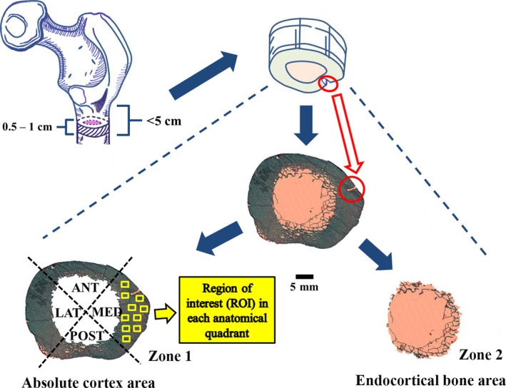
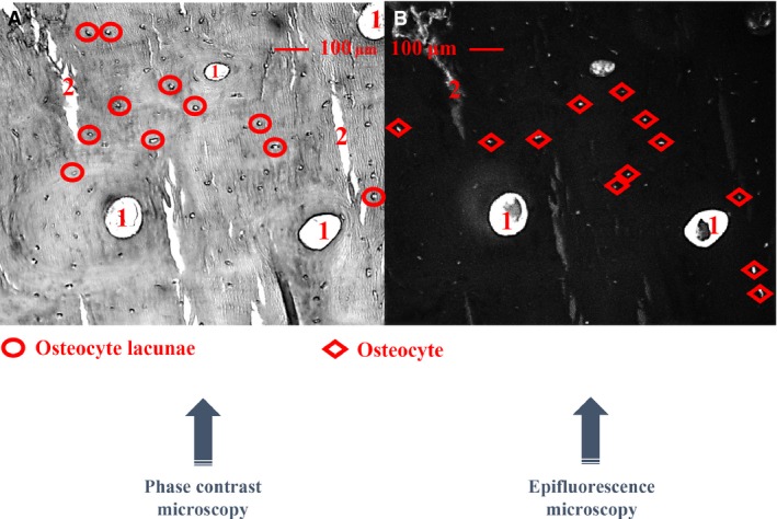
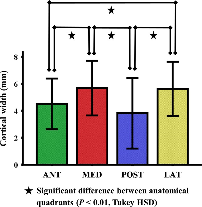
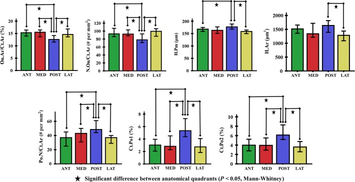
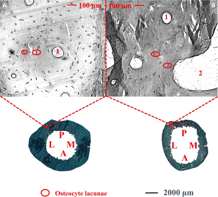
The histomorphometric properties of the subtrochanteric femoral region have rarely been investigated. The aim of this study was to investigate the age-associated variations and regional differences of histomorphometric and osteocytic properties in the cortical bone of the subtrochanteric femoral shaft, and the association between osteocytic and histological cortical bone parameters. Undecalcified histological sections of the subtrochanteric femoral shaft were obtained from cadavers (n = 20, aged 18-82 years, males). They were cut and stained using modified Masson-Goldner stain. Histomorphometric parameters of cortical bone were analysed with ×50 and ×100 magnification after identifying cortical bone boundaries using our previously validated method. Within cortical bone areas, only complete osteons with typical concentric lamellae and cement line were selected and measured. Osteocytic parameters of cortical bone were analyzed under phase contrast microscopy and epifluorescence within microscopic fields (0.55 mm2 for each). The cortical widths of the medial and lateral quadrants were significantly higher than other quadrants (P < 0.01). Osteonal area per cortical bone area was lower and cortical porosities were higher in the posterior quadrant than in the other quadrants (P < 0.05). Osteocyte lacunar number per cortical bone area was found higher in the young subjects (≤ 50 years) than in the older ones (> 50 years) both before and after adjustments for body height and weight (P < 0.05). Moreover, significant but low correlations were found between the cortical bone and osteocytic parameters (0.20 ≤ R2 ≤ 0.35, P < 0.05). It can be concluded that in healthy males, the cortical histomorphometric parameters differ between the anatomical regions of the subtrochanteric femoral shaft, and are correlated with the osteocytic parameters from the same site. These findings may be of use when discussing mechanisms that predispose patients to decreasing bone strength.
Keywords: bone histomorphometry; cortical bone; osteocyte; regional difference; subtrochanteric femoral shaft.
© 2017 Anatomical Society.
Figures
References
-
- Baumgaertner MR, Curtin SL, Lindskog DM, et al. (1995) The value of the tip‐apex distance in predicting failure of fixation of peritrochanteric fractures of the hip. J Bone Joint Surg Am 77, 1058–1064. - PubMed
-
- Beck TJ, Looker AC, Ruff CB, et al. (2000) Structural trends in the aging femoral neck and proximal shaft: analysis of the Third National Health and Nutrition Examination Survey dual‐energy X‐ray absorptiometry data. J Bone Miner Res 15, 2297–2304. - PubMed
-
- Bell KL, Loveridge N, Power J, et al. (1999) Regional differences in cortical porosity in the fractured femoral neck. Bone 24, 57–64. - PubMed
-
- Bell KL, Loveridge N, Jordan GR, et al. (2000) A novel mechanism for induction of increased cortical porosity in cases of intracapsular hip fracture. Bone 27, 297–304. - PubMed
-
- Bernhard A, Milovanovic P, Zimmermann EA, et al. (2013) Micro‐morphological properties of osteons reveal changes in cortical bone stability during aging, osteoporosis, and bisphosphonate treatment in women. Osteoporos Int 24, 2671‐80. - PubMed
MeSH terms
LinkOut - more resources
Full Text Sources
Other Literature Sources
