. 2017 Oct 15;595(20):6569-6583.
doi: 10.1113/JP274667. Epub 2017 Sep 13.

Median preoptic glutamatergic neurons promote thermoregulatory heat loss and water consumption in mice

Affiliations

Median preoptic glutamatergic neurons promote thermoregulatory heat loss and water consumption in mice

Stephen B G Abbott et al. J Physiol. .

Abstract

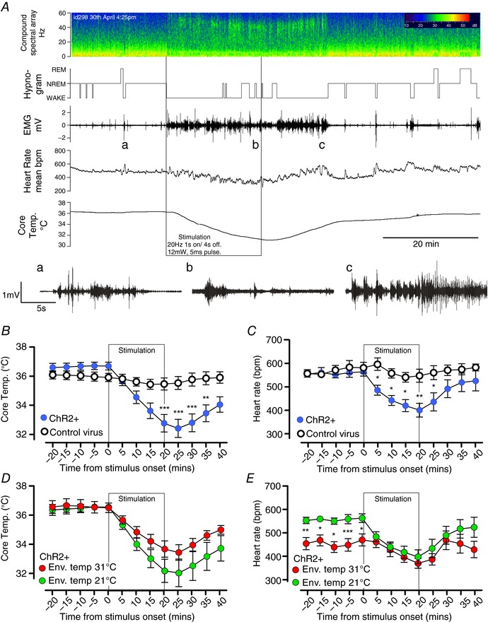
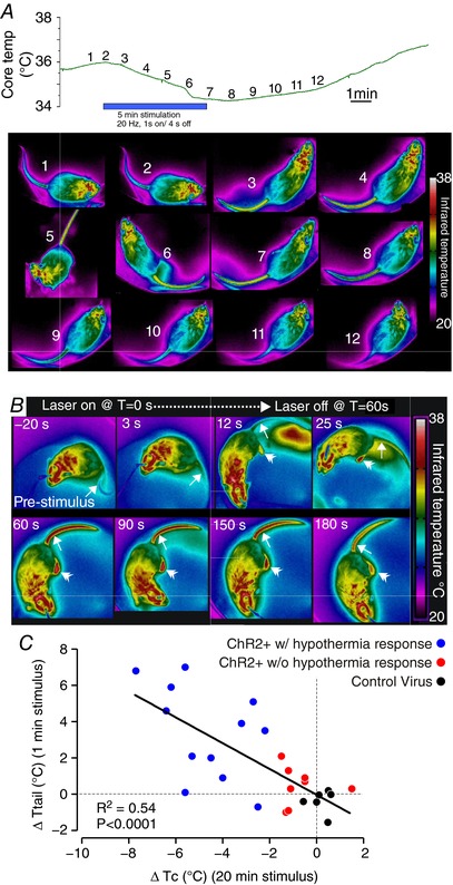
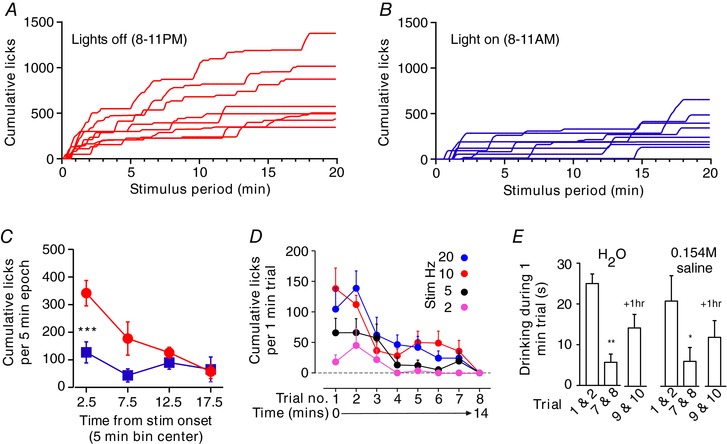
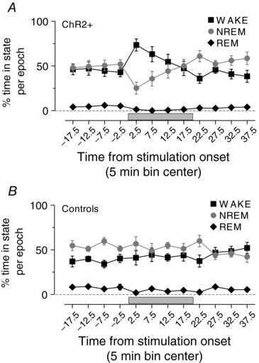
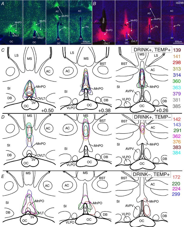
Key points: Glutamatergic neurons in the median preoptic area were stimulated using genetically targeted Channelrhodopsin 2 in transgenic mice. Stimulation of glutamatergic median preoptic area neurons produced a profound hypothermia due to cutaneous vasodilatation. Stimulation also produced drinking behaviour that was inhibited as water was ingested, suggesting pre-systemic feedback gating of drinking. Anatomical mapping of the stimulation sites showed that sites associated with hypothermia were more anteroventral than those associated with drinking, although there was substantial overlap.

Abstract: The median preoptic nucleus (MnPO) serves an important role in the integration of water/electrolyte homeostasis and thermoregulation, but we have a limited understanding these functions at a cellular level. Using Cre-Lox genetic targeting of Channelrhodospin 2 in VGluT2 transgenic mice, we examined the effect of glutamatergic MnPO neuron stimulation in freely behaving mice while monitoring drinking behaviour and core temperature. Stimulation produced a strong hypothermic response in 62% (13/21) of mice (core temperature: -4.6 ± 0.5°C, P = 0.001 vs. controls) caused by cutaneous vasodilatation. Stimulating glutamatergic MnPO neurons also produced robust drinking behaviour in 82% (18/22) of mice. Mice that drank during stimulation consumed 912 ± 163 μl (n = 8) during a 20 min trial in the dark phase, but markedly less during the light phase (421 ± 83 μl, P = 0.0025). Also, drinking during stimulation was inhibited as water was ingested, suggesting pre-systemic feedback gating of drinking. Both hypothermia and drinking during stimulation occurred in 50% of mice tested. Anatomical mapping of the stimulation sites showed that sites associated with hypothermia were more anteroventral than those associated with drinking, although there was substantial overlap. Thus, activation of separate but overlapping populations of glutamatergic MnPO neurons produces effects on drinking and autonomic thermoregulatory mechanisms, providing a structural basis for their frequently being coordinated (e.g. during hyperthermia).

Keywords: autonomic nervous system; hypothalamus; hypothermia; optogenetic; sympathetic nervous system; temperature; thermoregulation; thirst.


Figures

Figure 1
Figure 1. Time course of the effects of glutamatergic MnPO neuron stimulation
A, experimental recording of changes in EEG compound spectral activity, wake–sleep state (hypnogram), neck muscle activity (EMG), heart rate (HR) and core temperature (T c) in response to 20 min of phasic optogenetic stimulation of MnPO glutamatergic neurons (mouse id 298). Note that the animal awakens almost immediately with onset of stimulation, but that HR and T c gradually fall throughout the stimulation period. Insets a–c: expanded EMG trace before, during and after stimulation; note the rhythmic bursts in EMG representing shivering in c. B and C, grouped data of T c (B) and HR (C) changes during stimulation of VGlut2‐ires‐CreChR2+ mice (n = 14) with a hypothermic response and controls (n = 7). D and E, grouped data of T c (D) and heart rate (E) changes during stimulation in the light phase in VGlut2‐ires‐CreChR2+ (n = 7) with a hypothermic response in room temperature conditions (nominally 21°C) and thermoneutral (nominally 31°C) conditions. *** P < 0.001, ** P < 0.01, ** P < 0.05, t test with Bonferroni correction.
Figure 2
Figure 2. Stimulation causes heat loss through rapid vasodilatation of the tail
A, time course of changes in T c (upper panel) and tail temperature (T tail, lower panel, one photo each minute at the time shown on the upper panel) during 5 min of phasic stimulation. Note that increasing T tail (arrow) precedes a decrease in T c following the beginning of the stimulus, and T tail decreases before T c recovers post‐stimulus. B, example thermographs used for analysis of T tail (single arrowhead) during a 60 s stimulus starting at T = 0. Note that temperature of the feet (double arrowhead) also increases during stimulation. C, x–y plot of ΔT c vs. ΔT tail in 20 VGluT2‐ires‐CreChR2+ and 6 mice injected with a control virus.
Figure 3
Figure 3. Drinking during MnPO stimulation is regulated by diurnal feed‐forward and inhibitory feedback mechanisms
A and B, individual time course of cumulative water spout licks during 20 min trials in 8 mice conducted in the dark (A, red) and light (B, blue) phase. C, distribution of cumulative licks in light and dark phase trials for cases in A and B. *** P < 0.001 t test with Bonferroni correction. D, spout licks during each of 8 trials performed over 15 min (n = 7) using different stimulation frequencies. E, time spent drinking distilled water or 0.154 m saline during initial trials (average of trials 1 and 2) and last trials (average of trials 7 and 8) of a series of 8 trials, and two trials performed after 1 h of recovery (average of trials 9 and 10). n = 5, * P < 0.05, ** P < 0.01 vs. trials 1 and 2, t test with Bonferroni correction.
Figure 4
Figure 4. MnPO stimulation promotes wakefulness
Grouped data of sleep/wake profile during stimulation in VGlut2‐ires‐Cre ChR2+ (A) and controls (B). Note that stimulations during the day and night are combined in this analysis.
Figure 5
Figure 5. Distribution of ChR2‐mCherry neurons in animals with specific physiological responses
A and B, sequential coronal sections of the median preoptic nucleus in a Slc17a6‐cre;Rpl10‐GFP (Vglut2‐GFP) mouse (A) and a VGlut2‐ires‐CreChR2+ mouse (id 298, B), showing typical placement of an injection and the optical fibre. Scale bar: 500 μm. CE, mapping of transduced neurons (irregular outlines) within 1 mm below the tip of the optical fibre for 10 VGlut2‐ires‐CreChR2+ mice with a hypothermic response and water consumption during stimulation (C), six with water consumption, but little or no T c change during stimulation (D), and four with T c reductions and no water consumption during stimulation (E). Abbreviations: 3V, 3rd ventricle; AC, anterior commissure; AVPV, anteroventral periventricular preoptic nucleus; BST, bed nucleus of the striatum; DB, nucleus of the diagonal band; LS, lateral septum; MS, medial septum; oc, optic chiasm; Pe, periventricular preoptic nucleus; SI, substantia innominate.

Similar articles

Cited by

References

    1. Abbott SB, Machado NL, Geerling JC & Saper CB (2016). Reciprocal control of drinking behavior by median preoptic neurons in mice. J Neurosci 36, 8228–8237. - PMC - PubMed
    1. Baker MA, Doris PA & Hawkins MJ (1983). Effect of dehydration and hyperosmolality on thermoregulatory water losses in exercising dogs. Am J Physiol Regul Integr Comp Physiol 244, R516–R521. - PubMed
    1. Cunningham JT, Beltz T, Johnson RF & Johnson AK (1992). The effects of ibotenate lesions of the median preoptic nucleus on experimentally‐induced and circadian drinking behavior in rats. Brain Res 580, 325–330. - PubMed
    1. Davern PJ & McKinley MJ (2013). Brain regions influenced by the lateral parabrachial nucleus in angiotensin II‐induced water intake. Neuroscience 252, 410–419. - PubMed
    1. Dimitrov EL, Kim YY & Usdin TB (2011). Regulation of hypothalamic signaling by tuberoinfundibular peptide of 39 residues is critical for the response to cold: a novel peptidergic mechanism of thermoregulation. J Neurosci 31, 18166–18179. - PMC - PubMed

Publication types