Review
. 2017 Oct;30(5):393-404.
doi: 10.1177/1971400917697503. Epub 2017 Mar 20.

Back pain and scoliosis in children: When to image, what to consider

Affiliations
Review

Back pain and scoliosis in children: When to image, what to consider

Sonia F Calloni et al. Neuroradiol J. 2017 Oct.

Abstract

Back pain and scoliosis in children most commonly present as benign and self-limited entities. However, persistent back pain and/or progressive scoliosis should always be taken seriously in children. Dedicated diagnostic work-up should exclude etiologies that may result in significant morbidity. Clinical evaluation and management require a comprehensive history and physical and neurological examination. A correct imaging approach is important to define a clear diagnosis and should be reserved for children with persistent symptoms or concerning clinical and laboratory findings. This article reviews the role of different imaging techniques in the diagnostic approach to back pain and scoliosis, and offers a comprehensive review of the main imaging findings associated with common and uncommon causes of back pain and scoliosis in the pediatric population.

Keywords: Back pain; CT; MR; imaging; infectious diseases; radiographs; scoliosis; spinal tumors; spine; spine dysraphism; spondylolysis; trauma.


Figures
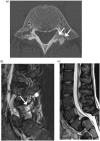
Figure 1.
Figure 1.
Spondylolysis. (a) Axial CT image shows a fracture/defect of the left pars interarticularis of a lumbar vertebra. (b) Sagittal T2-weighted images with fat saturation (STIR) magnetic resonance (MR) image shows hyperintense signal in the adjacent pedicle, consistent with bone-marrow edema from bone stress reaction. The fracture is seen as a hypointense line (arrow). (c) STIR image of a different patient with bilateral pars interarticularis fractures shows high-grade anterolisthesis of L5 on S1.
Figure 2.
Figure 2.
Viral myelitis secondary to enterovirus D68 infection. (a) Sagittal STIR MR image shows mild hyperintense signal and minimal swelling of the conus medullaris (arrows). (b) Axial T2-weighted MR image confirms the presence of abnormal hyperintense signal involving the bilateral anterior horns.
Figure 3.
Figure 3.
Drop metastasis. (a) Sagittal contrast enhanced T1-weighted MR image shows mildly enhancing nodular lesions anterior to the spinal cord (arrows). (b) Axial contrast enhanced T1-weighted image shows the intradural extramedullary lesion (arrow).
Figure 4.
Figure 4.
Astrocytoma. (a) Sagittal STIR MR image shows a heterogeneous infiltrative intramedullary mass expanding the thoracic spinal cord. Hyperintense signal at the cranial and caudal lesion may represent vasogenic edema or infiltrative tumor. (b) Sagittal fat-suppressed contrast enhanced MR image shows areas of nodular enhancement within the mass and posterior soft tissue postoperative changes in this child with recurrent tumor.
Figure 5.
Figure 5.
Ependymoma. Sagittal fat-suppressed contrast enhanced T1-weighted MR image shows heterogeneous mildly enhancing intraspinal mass involving the cervical and upper thoracic spinal cord.
Figure 6.
Figure 6.
Hemangioblastoma. (a) Sagittal T2-weighted image shows a round hypointense intramedullary lesion in the proximal thoracic spinal cord and extensive spinal cord edema. (b) Sagittal post-contrast T1-weighted image shows homogeneous intense enhancement of the lesion. Dilated vessels are seen in close proximity to this highly vascular tumor.
Figure 7.
Figure 7.
Langerhans cell histiocytosis. (a) Coronal computed tomography (CT) image shows a C4 lytic lesion with areas of cortical disruption and loss of vertebral body height. (b) Sagittal contrast enhanced T1-weighted MR image shows homogenous enhancement and anterior epidural extension.
Figure 8.
Figure 8.
Neurofibroma. (a) Sagittal T2-weighted image shows a confluent hyperintense extradural and paraspinal mass expanding several cervical neural foramina. (b) Axial T2-weighted MR image shows compression of the spinal canal and enlargement of the neural foramina by the bilateral extradural masses.
Figure 9.
Figure 9.
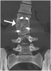
Transitional lumbosacral anatomy. Frontal view of a volume rendered image shows a large left L5 transverse process articulating with the sacrum (arrow), as well as left convex lumbar scoliosis. Notice the hardware fixating the pubic symphysis diastasis in this patient with history of bladder exstrophy.
Figure 10.
Figure 10.
Syringohydromyelia. Sagittal T1-weighted MR image shows marked cystic dilatation of the spinal cord in this child with Chiari I deformity.
Figure 11.
Figure 11.
Hemivertebra. Coronal CT image shows hemivertebra on the right side of L3 body (arrow) associated with focal right convex scoliosis of the lumbar spine.
Figure 12.
Figure 12.
Diastematomyelia. (a) Axial T2-weighted MR image shows an osseous septum splitting the spinal canal at L4–L5. (b) Sagittal T2-weighted MR image shows segmentation anomaly with fusion of the L4 and L5 vertebral bodies, as well as hydromyelia of the distal spinal cord.

References

    1. Altaf F, Heran MK, Wilson LF. Back pain in children and adolescents. Bone Joint J 2014; 96-B: 717–723. - PubMed
    1. Slipman CW, Patel RK, Botwin K, et al. epidemiology of spine tumors presenting to musculoskeletal physiatrists. Arch Phys Med Rehabil 2003; 84: 492–495. - PubMed
    1. Bernstein RM, Cozen H. Evaluation of back pain in children and adolescents. Am Fam Physician 2007; 76: 1669–1676. - PubMed
    1. Kjaer P, Leboeuf-Yde C, Sorensen JS, et al. An epidemiologic study of MRI and low back pain in 13-year-old children. Spine 2005; 30: 798–806. - PubMed
    1. Jackson C, McLaughlin K, Teti B. Back pain in children: a holistic approach to diagnosis and management. J Pediatr Health Care 2011; 25: 284–293. - PubMed

LinkOut - more resources