Sensitivity of docetaxel-resistant MCF-7 breast cancer cells to microtubule-destabilizing agents including vinca alkaloids and colchicine-site binding agents
- PMID: 28787019
- PMCID: PMC5546696
- DOI: 10.1371/journal.pone.0182400
Sensitivity of docetaxel-resistant MCF-7 breast cancer cells to microtubule-destabilizing agents including vinca alkaloids and colchicine-site binding agents
Abstract
Introduction: One of the main reasons for disease recurrence in the curative breast cancer treatment setting is the development of drug resistance. Microtubule targeted agents (MTAs) are among the most commonly used drugs for the treatment of breaset cancer and therefore overcoming taxane resistance is of primary clinical importance. Our group has previously demonstrated that the microtubule dynamics of docetaxel-resistant MCF-7TXT cells are insensitivity to docetaxel due to the distinct expression profiles of β-tubulin isotypes in addition to the high expression of p-glycoprotein (ABCB1). In the present investigation we examined whether taxane-resistant breast cancer cells are more sensitive to microtubule destabilizing agents including vinca alkaloids and colchicine-site binding agents (CSBAs) than the non-resistant cells.
Methods: Two isogenic MCF-7 breast cancer cell lines were selected for resistance to docetaxel (MCF-7TXT) and the wild type parental cell line (MCF-7CC) to examine if taxane-resistant breast cancer cells are sensitive to microtubule-destabilizing agents including vinca alkaloids and CSBAs. Cytotoxicity assays, immunoblotting, indirect immunofluorescence and live imaging were used to study drug resistance, apoptosis, mitotic arrest, microtubule formation, and microtubule dynamics.
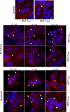
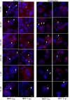
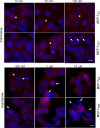
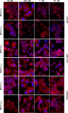
Results: MCF-7TXT cells were demonstrated to be cross resistant to vinca alkaloids, but were more sensitive to treatment with colchicine compared to parental non-resistant MCF-7CC cells. Cytotoxicity assays indicated that the IC50 of MCF-7TXT cell to vinorelbine and vinblastine was more than 6 and 3 times higher, respectively, than that of MCF-7CC cells. By contrast, the IC50 of MCF-7TXT cell for colchincine was 4 times lower than that of MCF-7CC cells. Indirect immunofluorescence showed that all MTAs induced the disorganization of microtubules and the chromatin morphology and interestingly each with a unique pattern. In terms of microtubule and chromain morphology, MCF-7TXT cells were more resistant to vinorelbine and vinblastine, but more sensitive to colchicine compared to MCF-7CC cells. PARP cleavage assay further demonstrated that all of the MTAs induced apoptosis of the MCF-7 cells. However, again, MCF-7TXT cells were more resistant to vinorelbine and vinblastine, and more sensitive to colchicine compared to MCF-7CC cells. Live imaging demonstrated that the microtubule dynamics of MCF-7TXT cells were less sensitive to vinca alkaloids, and more sensitive to colchicine. MCF-7TXT cells were also noted to be more sensitive to other CSBAs including 2MeOE2, ABT-751 and phosphorylated combretastatin A-4 (CA-4P).
Conclusion: Docetaxel-resistant MCF-7TXT cells have demonstrated cross-resistance to vinca alkaloids, but appear to be more sensitive to CSBAs (colchicine, 2MeOE2, ABT-751 and CA-4P) compared to non-resistant MCF-7CC cells. Taken together these results suggest that CSBAs should be evaluated further in the treatment of taxane resistant breast cancer.
Conflict of interest statement
Figures


References
-
- Lal S, Mahajan A, Chen WN, Chowbay B. Pharmacogenetics of target genes across doxorubicin disposition pathway: a review. CurrDrug Metab. 2010;11(1):115–28. - PubMed
-
- Jemal A, Bray F, Center MM, Ferlay J, Ward E, Forman D. Global cancer statistics. CA Cancer JClin. 2011;61(2):69–90. - PubMed
-
- Murray S, Briasoulis E, Linardou H, Bafaloukos D, Papadimitriou C. Taxane resistance in breast cancer: mechanisms, predictive biomarkers and circumvention strategies. Cancer TreatRev. 2012;38(7):890–903. - PubMed
-
- Kamangar F, Dores GM, Anderson WF. Patterns of cancer incidence, mortality, and prevalence across five continents: defining priorities to reduce cancer disparities in different geographic regions of the world. Journal of clinical oncology: official journal of the American Society of Clinical Oncology. 2006;24(14):2137–50. Epub 2006/05/10. doi: 10.1200/jco.2005.05.2308. . - DOI - PubMed
-
- Yardley DA. Drug resistance and the role of combination chemotherapy in improving patient outcomes. Int J Breast Cancer. 2013;2013:137414 Epub 2013/07/19. doi: 10.1155/2013/137414. ; PubMed Central PMCID: PMCPmc3707274. - DOI - PMC - PubMed
MeSH terms
Substances
LinkOut - more resources
Full Text Sources
Other Literature Sources
Medical

