. 2017 Oct;106(2):236-251.
doi: 10.1111/mmi.13760. Epub 2017 Aug 22.

Carbonyl reduction by YmfI in Bacillus subtilis prevents accumulation of an inhibitory EF-P modification state

Affiliations

Carbonyl reduction by YmfI in Bacillus subtilis prevents accumulation of an inhibitory EF-P modification state

Katherine R Hummels et al. Mol Microbiol. 2017 Oct.

Abstract

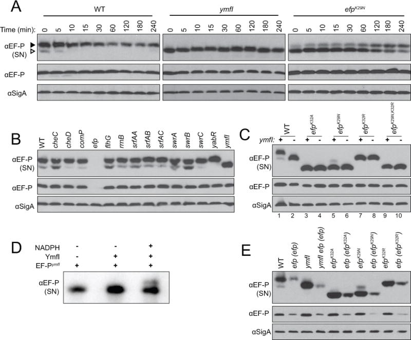
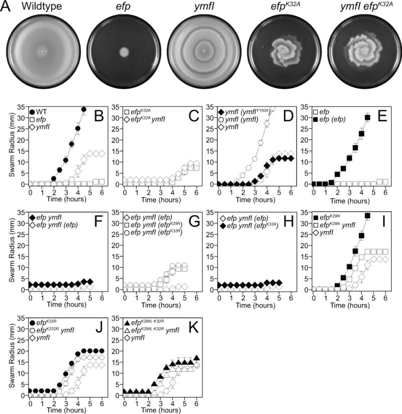
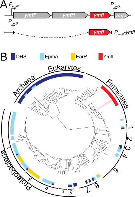
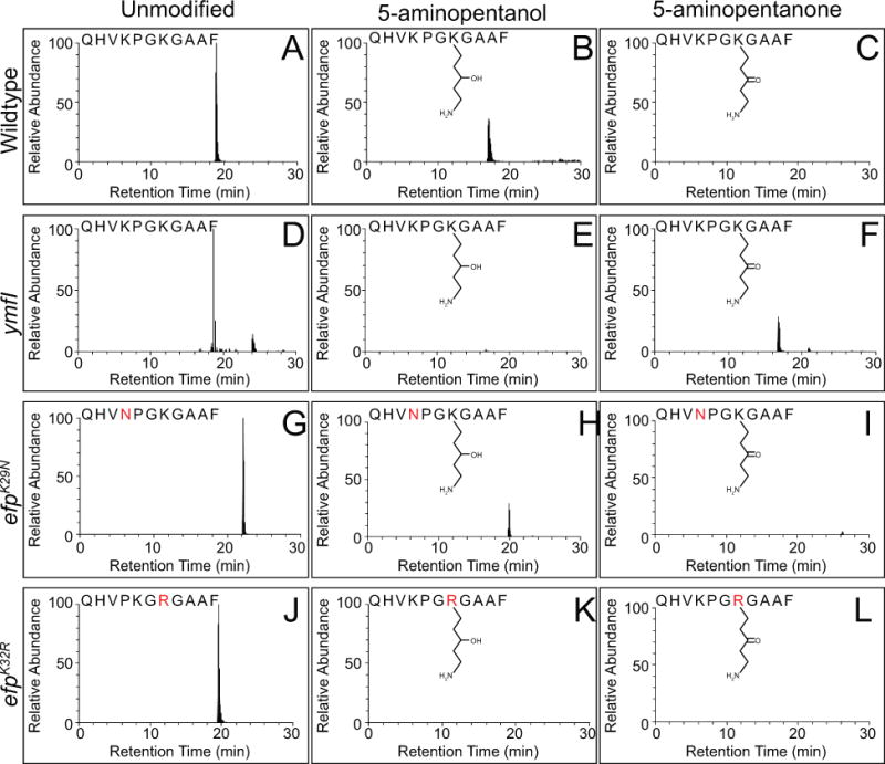
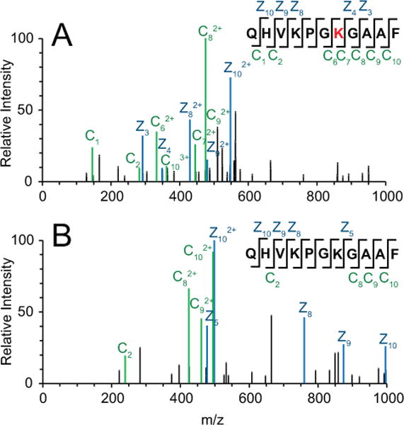
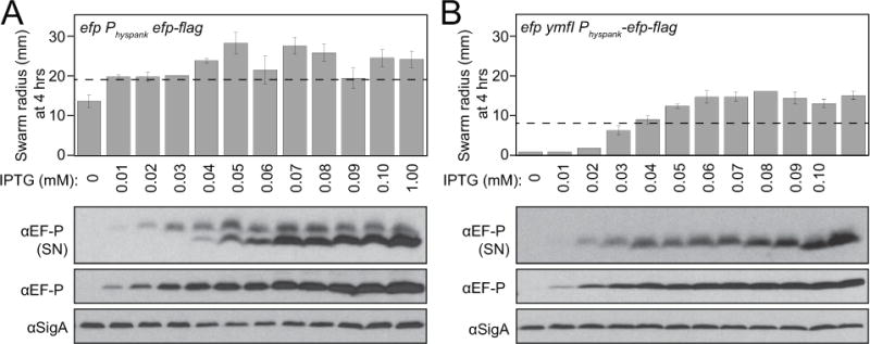
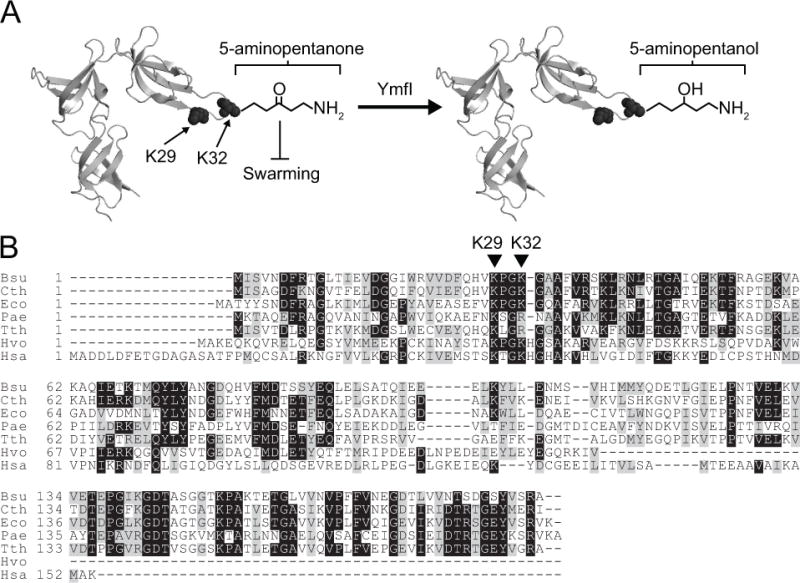
Translation elongation factor P (EF-P) in Bacillus subtilis is required for a form of surface migration called swarming motility. Furthermore, B. subtilis EF-P is post-translationally modified with a 5-aminopentanol group but the pathway necessary for the synthesis and ligation of the modification is unknown. Here we determine that the protein YmfI catalyzes the reduction of EF-P-5 aminopentanone to EF-P-5 aminopentanol. In the absence of YmfI, accumulation of 5-aminopentanonated EF-P is inhibitory to swarming motility. Suppressor mutations that enhanced swarming in the absence of YmfI were found at two positions on EF-P, including one that changed the conserved modification site (Lys 32) and abolished post-translational modification. Thus, while modification of EF-P is thought to be essential for EF-P activity, here we show that in some cases it can be dispensable. YmfI is the first protein identified in the pathway leading to EF-P modification in B. subtilis, and B. subtilis encodes the first EF-P ortholog that retains function in the absence of modification.


Figures

Figure 1
Figure 1. EF-P resolves as two species on a semi-native gel in a YmfI-dependent manner
(A) Translation was inhibited in mid-log phase cultures by the addition of spectinomycin, lysates were subsequently harvested at the indicated time points, resolved by semi-native (top panel, “SN”) or denaturing (middle and bottom panels) polyacrylamide gel electrophoresis, electroblotted, and probed with anti-EF-P or anti-SigA polyclonal antisera (used as a loading control) as indicated. The following strains were used to generate the samples: wild type (DK1042), ymfI (DK3621), and efpK29N (DK4282). For denaturing gels, EF-P resolved at approximately 22 kDa and SigA resolved at approximately 46 kDa. The black arrow indicates the upper band and the white arrow indicates the lower band. (B) Lysates of mid-log phase cultures were resolved by semi-native (top panel, “SN”) or denaturing (middle and bottom panels) polyacrylamide gel electrophoresis, electroblotted, and probed with anti-EF-P or anti-SigA polyclonal antisera as indicated. The following strains were used to generate the samples: wild type (3610), cheC (DS1045), cheD (DS1064), comP (DS1028), efp (DS1124), flhG (DS1164), rrnB-16S (DS1146), srfAA (DS1102), srfAB (DS1044), srfAC (DS1122), swrA (DS1026), swrB (DS1107), swrC (DS1113), yabR (DS1078), and ymfI (DS1029) (C) Lysates of mid-log phase cultures were resolved by semi-native (top panel, “SN”) or denaturing (middle and bottom panels) polyacrylamide gel electrophoresis, electroblotted, and probed with anti-EF-P or anti-SigA polyclonal antisera as indicated. Lanes are numbered at the bottom of the panels for clarity in text. The presence and absence of a wild type copy of the ymfI gene is indicated by (+) and (-) respectively at the top of the panels. The following strains were used to generate samples: wild type (DK1042), ymfI (DK3621), efpK32A (DK3235), efpK32A ymfI (DK3712), efpK29N (DK4282), efpK29N ymfI (DK4396), efpK32R (DK4359), efpK32R ymfI (DK4397), efpK29N,K32R (DK4420), and efpK32A,K32R ymfI (DK4436). (D) EF-P-FLAG purified from a ymfI mutant (EF-P-FLAGymfI) was incubated alone, with YmfI, or with YmfI and 150 mM NADPH for 30 min at 37C. Reactions were subsequently resolved by semi-native gel electrophoresis, electroblotted, and probed with anti-EF-P antisera. (E) Lysates of mid-log phase cultures were resolved by semi-native (top panel, “SN”) or denaturing (middle and bottom panels) polyacrylamide gel electrophoresis, electroblotted, and probed with anti-EF-P or anti-SigA polyclonal antisera as indicated. The following strains were used to generate the samples: wild type (DK1042), efp (efp) (DK3780), ymfI (DK3621), ymfI efp (efp) (DK3789), efpK32A (DK3235), efp (efpK32A) (DK2248), efpK29N (DK4282), efp (efpK29N) (DK4043), efpK32R (DK4359), efp (efpK32R) (DK4072).
Figure 2
Figure 2. Cells mutated for ymfI are defective in swarming motility and swarming can be restored by mutations in efp
(A) Top views of centrally-inoculated swarm plates incubated overnight at 37°C were imaged against a black background. Zones of colonization appear light grey. The plate inoculated with the ymfI mutant has internal rings that mark the locations at which the population stopped moving and restarted at a later time point. Note: a comparable cessation can be seen at hour 5 in the quantitative swarm expansion assay in panel 2B. The following strains were used to generate the panels: wild type (DK1042), efp (DK2050), ymfI (DK3621), efpK32A (DK3235), and ymfI efpK32A (DK3712).(B-K) Quantitative swarm expansion assays in which mid-log phase cultures were concentrated and used to inoculate swarm plates. Swarm expansion was monitored along the same axis every 30 min for 5–6 hours. Each data point represents the average of three replicates and error bars represent the standard deviation. The following strains were used as the inoculum (B) wild type (DK1042), efp (DK2050), and ymfI (DK3621). (C) efpK32A (DK3235) and efpK32A ymfI (DK3712). (D) ymfI (ymfIY150A) (DK4233), ymfI (ymfI) (DK3969), and ymfI (DK3621). (E) efp (DK2050) and efp (efp) (DK3780). (F) efp ymfI (DK2886), and efp ymfI (efp) (DK3789). (G) efp ymfI (efp) DK3789, efp ymfI (efpK29N) (DK4043), and, efp ymfI (efpK32R) (DK4072). (H) efp ymfI (efp) (DK3789) and efp ymfI (efpK32A) (DK2889). (I) efpK29N (DK4282), efpK29N ymfI (DK4396), and ymfI (DK3621). (J) efpK32R (DK4359), efpK32R ymfI (DK4397), and ymfI (DK3621). (K) efpK29N,K32R (DK4420), efpK29N,K32R ymfI (DK4436), and ymfI (DK3621).
Figure 3
Figure 3. Genetic architecture and phylogenetic distribution of the ymfI locus
(A) Predicted genetic architecture of the ymfI locus and the design of the PymfF-ymfI complementation construct. Both ymfF and ymfH are predicted to encode peptidases and ymfJ encodes a DUF3235-domain containing protein. (B) The distribution of homologs of the EF-P modification enzymes deoxyhypusine synthase, DHS (dark blue), EpmA (light blue), EarP (yellow), and YmfI (red) across the three domains of life. YmfI is a member of the large family of alcohol dehydrogenases and the proximity to neighboring genes as indicated in Fig 3A was used to aid in the identification of YmfI homologs. Where multiple enzymes are encoded in the same genome, the bar is split accordingly. White space indicates the absence of a homolog to any EF-P modification enzyme. Numbers indicate the following clades (1) Flavobacterium-Cytophaga-Bacteroides group, (2) Chlamydiales, (3) Planctomycetes, (4) Spirochaetes, (5) Actinobacteria, (6) Deinococcus-Thermus group, and (7) Cyanobacteria. Bacillus subtilis is highlighted in pink. See Fig S5 enlarged map with species names. Table S3 contains the accession numbers for each species and Table S4 contains all sequences called to be YmfI in all completed bacterial genomes to date.
Figure 4
Figure 4. Deletion of ymfI results in 5-aminopentanonation of EF-P and can be suppressed by abolishing EF-P post-translational modification
Extracted ion chromatograms of chymotrypsin-digested EF-P peptide from wild type (A–C), ymfI mutant (D–F), efpK29N mutant (G–I), and efpK32R mutant (J–L). Three different species were detected for the peptide corresponding to the wild type sequence QHVKPGKGAAF containing including unmodified (A,D,G,J), 5-aminopentanolylated (B,E,H,K), and 5-aminopentanonated (C,F,I,L) lysine residue 32. All chromatograms for ymfI represent the 2+ ion for the indicated peptide. All other chromatograms represent the 3+ ion for the indicated peptide. Point mutations are indicated in bold red lettering. Predicted chemical structures are indicated. The cartoon indicates that the hydroxyl/carbonyl group is at the C3 position but it is possible that the hydroxyl group is at the C4 position instead. The precise position of the carbonyl can only be unequivocally determined with the use NMR or other high resolution structural methods.
Figure 5
Figure 5. EF-P is 5-aminopentanonated in the absence of YmfI
MS2 spectrum generated from ETD fragmentation of the QHVKPGKGAAF peptide. Both (A) Unmodified (precursor m/z of 570.320) and (B) 5-aminopentanonated (precursor m/z of 413.752) peptide were detected in the absence of ymfI. z ions are indicated in blue, and c ions are indicated in green. Site of 5-aminopentanonation (Lys32) is marked in red.
Figure 6
Figure 6. Overexpression of EF-P results in hyper-accumulation of the unmodified form and partially bypasses the need for YmfI
A) An efp mutant and B) a efp ymfI double mutant each with an ectopically integrated Physpank-EF-P-FLAG construct was induced with the indicated concentrations of IPTG and measured for swarming motility radius after 4 hours of incubation (upper panel) and Western blot analysis following semi-native gel (SN) (top row) and denaturing (middle and bottom rows) electrophoresis of cell lystates (lower panel). Note, dashed line on 4 hr swarm expansion graph indicates the extent of expansion of either wild type (A) or a ymfI mutant (B). The following strains were used to generate samples: efp Physpank-efp-flag (DK2448) and efp ymfI Physpank-efp-flag (DK3828). Wild type (DK1042) and ymfI (DK3621) were used to generate the dashed lines.
Figure 7
Figure 7. Model of EF-P activation by YmfI
(A) The crystal structure of EF-P isolated from the close relative of B. subtilis, Clostridium thermocellum (PDB ID 1YBY) with the location of Lys 29 (K29) and Lys 32 (K32) indicated. YmfI catalyzes the converstion of 5-aminopentanone (left) to 5-aminopentanol (right) groups on Lys32. In the absence of YmfI, 5-aminopentanone accumulates on EF-P and inhibits swarming motility (T-bar). (B) A multiple sequence alignment of EF-P orthologs from B. subtilis (Bsu), C. thermocellum (Cth), E. coli (Eco), Pseudomonas aeruginosa (Pae), Thermus thermophilus (Tth), the archaeon Haloferax volcanii (Hvo), and the eukaryote Homo sapiens (Hsa). The location of B. subtilis EF-P conserved residues K29 and K32 are indicated by carets.

Similar articles

Cited by

References

    1. Balibar CJ, Iwanowicz D, Dean CR. Elongation Factor P is dispensable in Escherichia coli and Pseudomonas aeruginosa. Curr Microbiol. 2013;67:293–299. - PubMed
    1. Bendezu FO, Hale CA, Bernhardt TG, de Boer PA. RodZ (YfgA) is required for proper assembly of MreB actin cytoskeleton and cell shape in E. coli. EMBO J. 2009;28:581–590. - PMC - PubMed
    1. Blaha G, Stanley RE, Steitz TA. Formation of the first peptide bond: the structure of EF-P bound to the 70S ribosome. Science. 2009;353:966–969. - PMC - PubMed
    1. Camacho C, Coulouris G, Avagyan V, Ma N, Papadopoulos J, Bealer K, Madden TL. BLAST+: acrchitecture and applications. BMC Bioinformatics. 2009;10:421. - PMC - PubMed
    1. Cooper HL, Park MH, Folk JE, Safer B, Braverman R. Identification of the hypusine-containing protein Hy+ as translation initiation factor eIF-4D. Proc Natl Acad Sci. 1983;80:1854–1857. - PMC - PubMed

MeSH terms

LinkOut - more resources