

Improved Resin-Zirconia Bonding by Room Temperature Hydrofluoric Acid Etching
- PMID: 28787975
- PMCID: PMC5455444
- DOI: 10.3390/ma8030850
Improved Resin-Zirconia Bonding by Room Temperature Hydrofluoric Acid Etching
Abstract
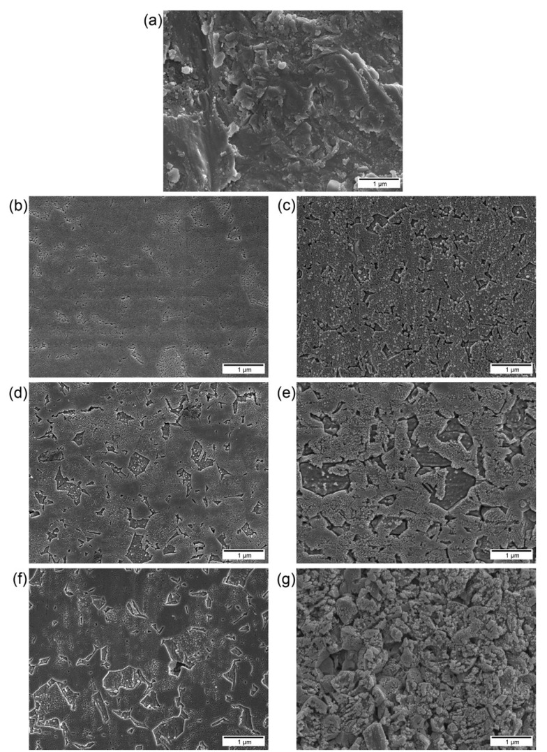
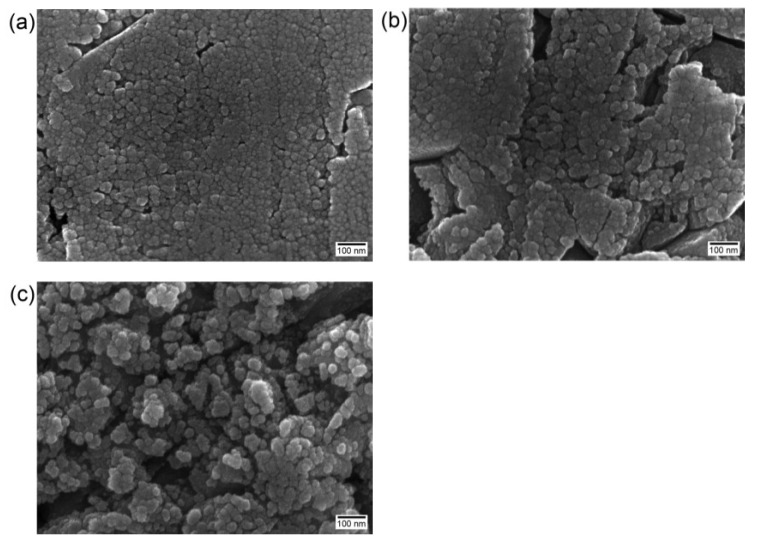
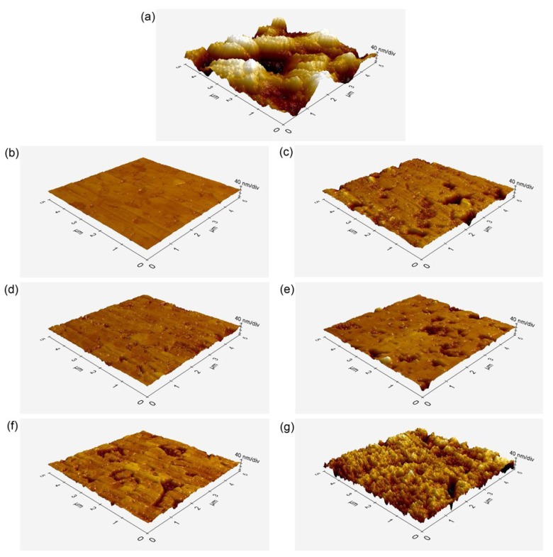
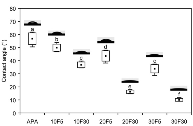
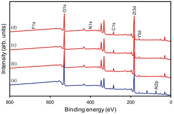
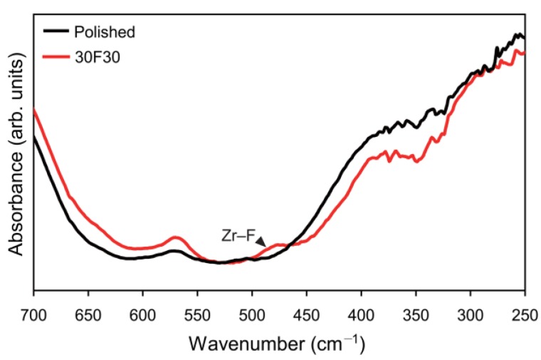
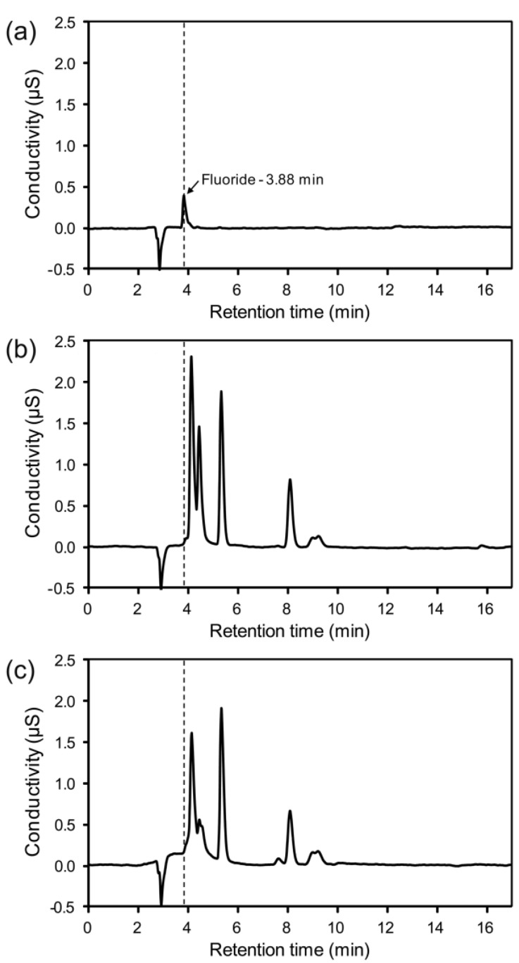
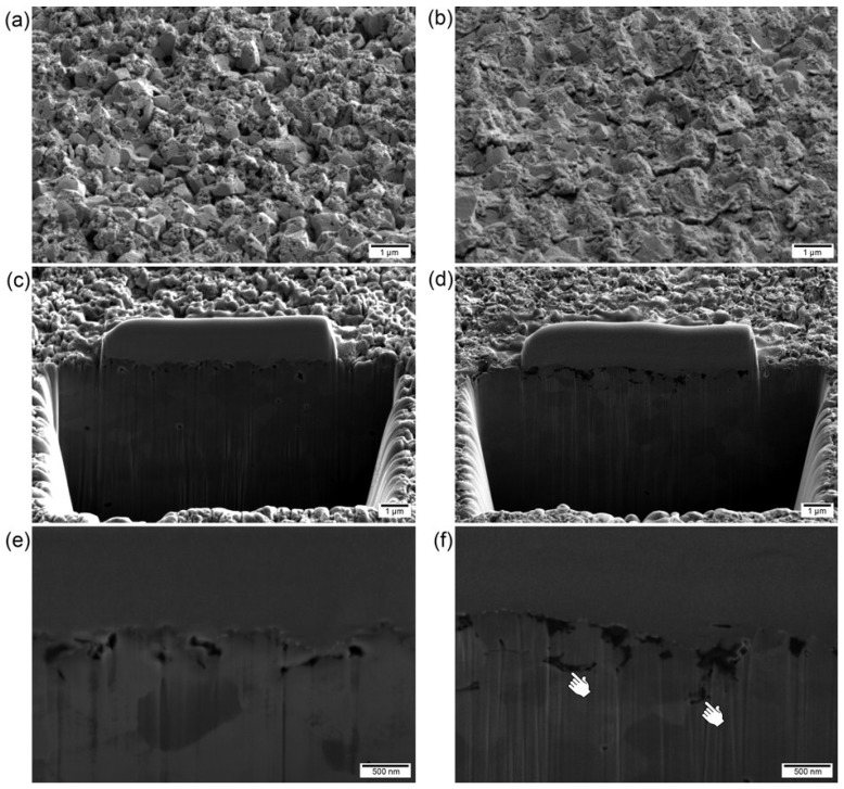
This in vitro study was conducted to evaluate the shear bond strength of "non-self-adhesive" resin to dental zirconia etched with hydrofluoric acid (HF) at room temperature and to compare it to that of air-abraded zirconia. Sintered zirconia plates were air-abraded (control) or etched with 10%, 20%, or 30% HF for either 5 or 30 min. After cleaning, the surfaces were characterized using various analytical techniques. Three resin cylinders (Duo-Link) were bonded to each treated plate. All bonded specimens were stored in water at 37 °C for 24 h, and then half of them were additionally thermocycled 5000 times prior to the shear bond-strength tests (n = 12). The formation of micro- and nano-porosities on the etched surfaces increased with increasing concentration and application time of the HF solution. The surface wettability of zirconia also increased with increasing surface roughness. Higher concentrations and longer application times of the HF solution produced higher bond-strength values. Infiltration of the resin into the micro- and nano-porosities was observed by scanning electron microscopy. This in vitro study suggests that HF slowly etches zirconia ceramic surfaces at room temperature, thereby improving the resin-zirconia bond strength by the formation of retentive sites.
Keywords: etching; hydrofluoric acid; resin–zirconia bonding; zirconia ceramic.
Conflict of interest statement
The authors declare no conflict of interest.
Figures
References
-
- Yun J.Y., Ha S.R., Lee J.B., Kim S.H. Effect of sandblasting and various metal primers on the shear bond strength of resin cement to Y-TZP ceramic. Dent. Mater. 2010;26:650–658. - PubMed
-
- Ha J.Y., Son J.S., Kim Y.K., Kim K.H., Kwon T.Y. Effect of heat treatment of dental zirconia ceramic treated with three different primers on the bonding durability of resin cement. Macromol. Res. 2013;21:71–77.
-
- Villard N., Seneviratne C., Tsoi J.K., Heinonen M., Matinlinna J. Candida albicans aspects of novel silane system-coated titanium and zirconia implant surfaces. Clin. Oral Implants Res. 2015;26:332–341. - PubMed
-
- Lohbauer U., Zipperle M., Rischka K., Petschelt A., Müller F.A. Hydroxylation of dental zirconia surfaces: Characterization and bonding potential. J. Biomed. Mater. Res. B Appl. Biomater. 2008;87:461–467. - PubMed
-
- Piascik J.R., Swift E.J., Braswell K., Stoner B.R. Surface fluorination of zirconia: Adhesive bond strength comparison to commercial primers. Dent. Mater. 2012;28:604–608. - PubMed
LinkOut - more resources
Full Text Sources
Other Literature Sources
Research Materials
Miscellaneous
