Review
. 2017 Jul 28;18(8):1406.
doi: 10.3390/ijms18081406.

Cellular Reparative Mechanisms of Mesenchymal Stem Cells for Retinal Diseases

Affiliations
Review

Cellular Reparative Mechanisms of Mesenchymal Stem Cells for Retinal Diseases

Suet Lee Shirley Ding et al. Int J Mol Sci. .

Abstract

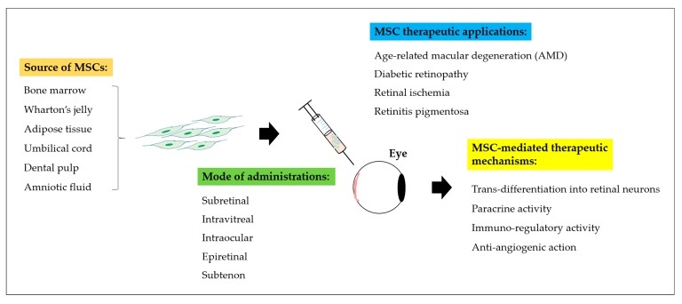
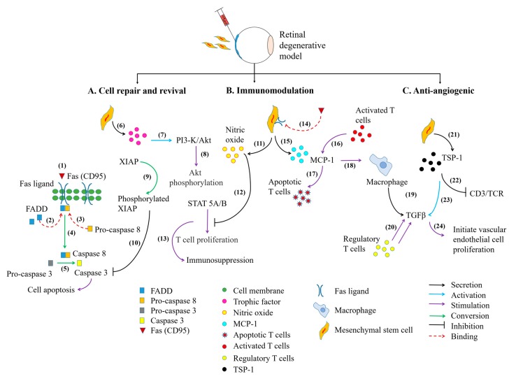
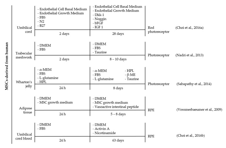
The use of multipotent mesenchymal stem cells (MSCs) has been reported as promising for the treatment of numerous degenerative disorders including the eye. In retinal degenerative diseases, MSCs exhibit the potential to regenerate into retinal neurons and retinal pigmented epithelial cells in both in vitro and in vivo studies. Delivery of MSCs was found to improve retinal morphology and function and delay retinal degeneration. In this review, we revisit the therapeutic role of MSCs in the diseased eye. Furthermore, we reveal the possible cellular mechanisms and identify the associated signaling pathways of MSCs in reversing the pathological conditions of various ocular disorders such as age-related macular degeneration (AMD), retinitis pigmentosa, diabetic retinopathy, and glaucoma. Current stem cell treatment can be dispensed as an independent cell treatment format or with the combination of other approaches. Hence, the improvement of the treatment strategy is largely subjected by our understanding of MSCs mechanism of action.

Keywords: MSC differentiation; anti-angiogenesis; anti-inflammatory; immunomodulatory; mesenchymal stem cells; paracrine activity; retinal degenerative diseases.


Conflict of interest statement

The authors declare no conflict of interest.

Figures

Figure 1
Figure 1
The basic retinal structure. Histological appearance of choroid and retinal layers. The retina is arranged in different layers of cells, from Retinal Pigment Epithelium (RPE), Outer Nuclear Layer (ONL), Outer Plexiform Layer (OPL), Inner Nuclear Layer (INL), Inner Plexiform Layer (IPL), and ganglion cell layer. The retinal layer harbors five retinal neuronal cells, primarily, the rod- and cone-photoreceptors, the Müller glia, the horizontal cell, the bipolar cell, the amacrine cell, and the Retinal Ganglion Cell (RGC). The arrow indicates the light transmission into the retina. Modified with permission from InTech’s Publishing Ethics and Legal Affairs Department [5] (© 2012 Triviño A, De Hoz R, Rojas B, Gallego BI, Ramírez AI, Salazar JJ, Ramírez JM. Published in [short citation] under CC BY 3.0 license. Available from: http://dx.doi.org/10.5772/48359).
Figure 2
Figure 2
A schematic representation of Mesenchymal Stem Cells (MSCs) therapeutic strategies in retinal degenerative diseases. Different sources of MSC such as bone marrow, Wharton’s jelly, adipose tissue, umbilical cord, dental pulp, and amniotic fluid have been discovered. Multiple routes of administration including subretinal, intravitreal, intraocular, epiretinal or subtenon injections can be implemented to deliver MSCs into the posterior lining of the eye. Delivery of MSCs into patients affected with posterior eye diseases including Age-related Macular Degeneration (AMD), diabetic retinopathy, retinal ischemia, and retinitis pigmentosa can be restored through trans-differentiation, paracrine activity, immuno-regulatory function, and anti-angiogenic action of MSCs.
Figure 3
Figure 3
The signaling pathways involved in MSC-mediated therapeutic strategies in the eye. The cell death machinery involves (12) the binding of Fas/Fas ligand, which assembles Fas-Associated protein with Death Domain (FADD) to form a docking site for pro-caspase 8. This event initiates the (34) crosslinking of pro-caspase 8 to FADD and activates caspase 8. Activated caspase 8 (5) induces the conversion of pro-caspase 3 into caspase 3 which are essential for the initiation of cell apoptosis. The MSCs cellular reparative action can be exerted (67) by the release of its beneficial trophic factors, including IL-6 which could further promote the migration of MSCs towards site of injury. The binding of IL-6 on MSCs will activate Phosphatidylinositol-3-Kinase (PI3-K)/Akt signaling pathway. (810) The phosphorylated Akt then induces X-linked Inhibitor of Apoptosis Protein (XIAP) phosphorylation leading to inhibition of caspase 3 activity. The immunomodulatory action of MSCs can be depicted through (1113) MSCs secretion of nitric oxide, which hampers Signal Transducer Activator-of-Transcription 5 (STAT5) phosphorylation and progressively leads to attenuation of T cell proliferation. The alleviation of T cell activity can be modulated through (1415) the expression of Fas ligand on MSC cell surface. This creates binding site for Fas protein, in which induces MSCs secretion of Monocyte Chemotactic Protein-1 (MCP-1) protein. (1617) The secreted proteins subsequently attract and induced apoptosis of activated T cells. (1820) Accumulation of apoptotic T cells further stimulate macrophage to release Transforming Growth Factor-beta (TGF-β) and subsequently recruit regulatory T cells. The regulatory T cells could also convert cytotoxic T cells into regulatory T cells. In addition, (2122) MSCs also secrete Thrombospondin type-1 (TSP-1) proteins to suppress Cluster of Differentiation 3 (CD3)/T cell receptor-mediated T cell proliferation. (2324) The released TSP-1 proteins activate TGF-β activity to initiate vascular endothelial cell remodeling. MSC cell differentiation is not mentioned in this figure.
Figure 4
Figure 4
A timeline representation of strategies used to direct human MSCs differentiation into retinal neurons and retinal pigmented epithelial cells, in vitro. Generation of retinal cell from MSCs involve the manipulation of stem cell fate by cytokines, growth factors, or inhibitory peptides. MSCs derived from different tissue origins including bone marrow, dental pulp, umbilical cord, trabecular meshwork, Wharton’s jelly, adipose tissue, and umbilical cord blood have previously demonstrated successful differentiation potential into retinal cells. The vast differentiation potential of MSCs into retinal cells includes photoreceptor, amacrine cell, RGC, and RPE-like cells that requires addition of specific cytokines, growth factors or inhibitory peptides. The differentiation factors are comprised of either taurine, activin A, basic Fibroblast Growth Factor (bFGF), β-mercaptoethanol (β-ME), Dickkopf Wnt signaling pathway inhibitor-1 (Dkk-1), noggin, insulin growth factor 1 (IGF 1), Human Platelet Lysate (HPL), and Vasoactive Intestinal Peptide. FBS, Fetal Bovine Serum; DMEM, Dulbecco’s Modified Eagle Medium; α-MEM, alpha Minimal Essential Medium; B27, B27 supplement; and N2, N2 supplement.

References

    1. Athanasiou D., Aguilà M., Bevilacqua D., Novoselov S.S., Parfitt D.A., Cheetham M.E. The cell stress machinery and retinal degeneration. FEBS Lett. 2013;587:2008–2017. doi: 10.1016/j.febslet.2013.05.020. - DOI - PMC - PubMed
    1. Mannu G.S. Retinal phototransduction. Neurosciences. 2014;19:275–280. - PMC - PubMed
    1. Reed B.T., Behar-Cohen F., Krantic S. Seeing early signs of Alzheimer’s Disease through the lens of the eye. Curr. Alzheimer Res. 2016;14:6–17. doi: 10.2174/1567205013666160819131904. - DOI - PubMed
    1. Palczewski K. Chemistry and biology of vision. J. Biol. Chem. 2012;287:1612–1619. doi: 10.1074/jbc.R111.301150. - DOI - PMC - PubMed
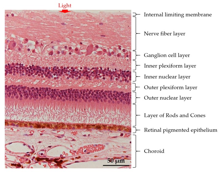
    1. Triviño A., de Hoz R., Rojas B., Gallego B.I., Ramírez A.I., Salazar J.J., Ramírez J.M. Ocular Diseases. InTech; Rijeka, Croatia: 2012. Effects of Hypercholesterolaemia in the Retina.

MeSH terms