Review
. 2014 Feb 10;7(2):980-1016.
doi: 10.3390/ma7020980.

Wear Debris Characterization and Corresponding Biological Response: Artificial Hip and Knee Joints

Affiliations
Review

Wear Debris Characterization and Corresponding Biological Response: Artificial Hip and Knee Joints

Md J Nine et al. Materials (Basel). .

Abstract

Wear debris, of deferent sizes, shapes and quantities, generated in artificial hip and knees is largely confined to the bone and joint interface. This debris interacts with periprosthetic tissue and may cause aseptic loosening. The purpose of this review is to summarize and collate findings of the recent demonstrations on debris characterization and their biological response that influences the occurrence in implant migration. A systematic review of peer-reviewed literature is performed, based on inclusion and exclusion criteria addressing mainly debris isolation, characterization, and biologic responses. Results show that debris characterization largely depends on their appropriate and accurate isolation protocol. The particles are found to be non-uniform in size and non-homogeneously distributed into the periprosthetic tissues. In addition, the sizes, shapes, and volumes of the particles are influenced by the types of joints, bearing geometry, material combination, and lubricant. Phagocytosis of wear debris is size dependent; high doses of submicron-sized particles induce significant level of secretion of bone resorbing factors. However, articles on wear debris from engineered surfaces (patterned and coated) are lacking. The findings suggest considering debris morphology as an important parameter to evaluate joint simulator and newly developed implant materials.

Keywords: biological response; isolation; morphology; nano-toxicity; wear debris.


Conflict of interest statement

The authors declare no conflict of interest.

Figures

Figure 1.
Figure 1.
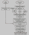
Flowchart illustrating the systematic search strategy of published peer-reviewed journals on wear-debris of hip and knee implants.
Figure 2.
Figure 2.
General method of Particle Isolation.
Figure 3.
Figure 3.
Typical morphologies of debris from joint simulator; (a) Carbon/Carbon composites [110]; and (b) CrCo alloy [111]; (c) Cylindrical (C/C composites) [110]; (d) Radial broken (C/C composites) [110]; (e) Blocky/Slice (C/C composites) [110]; (f) Fibril and Twig (UHMWPE) [34]; (g) Spherical (UHMWPE) [34] and (h) Sheet/flake type (UHMWPE) [34].
Figure 3.
Figure 3.
Typical morphologies of debris from joint simulator; (a) Carbon/Carbon composites [110]; and (b) CrCo alloy [111]; (c) Cylindrical (C/C composites) [110]; (d) Radial broken (C/C composites) [110]; (e) Blocky/Slice (C/C composites) [110]; (f) Fibril and Twig (UHMWPE) [34]; (g) Spherical (UHMWPE) [34] and (h) Sheet/flake type (UHMWPE) [34].
Figure 4.
Figure 4.
Typical morphologies of wear debris from periprosthetic tissue; (a) UHMWPE [90]; and (b) Alumina [103]; (c) Spherical (UHMWPE) [34]; (d) Sheet/Flake type (UlHMWPE) [112] and (e) Fibril (UHMWPE) [101].
Figure 5.
Figure 5.
AFM morphology of UHMWPE wear debris [126]; (a) A two-dimensional projection of AFM data for debris particles of the 0.2–0.8 μm fraction precipitated on a filter. Six of the larger particles and three pores are indicated; (b) three-dimensional projections of AFM data for the six particles indicated in pane (dimensions are in nm); and (c) examples of length (L), width (W), and height (H) measurements on two representative UHMWPE particles.
Figure 6.
Figure 6.
(a) TEM images of MG63 cells at 37 °C (incubation with Al2O3 NPs for 6 h), Arrows pointing to the process of internalization at the surface associated with actin rearrangement near the plasma membrane and extension into the extracellular space [150] and (b) SEM image of live primary human dermal fibroblasts exposed to CoCr alloy nanoparticles for 24 h outside and inside the cell [139].
Figure 7.
Figure 7.
(a) Saos-2 cells challenged for 24 h with 0.5 mg/mL of FeAlCr alloys (avg. dia. 3.7 ± 0.4) and (b) Mineral formation after 21 days by Saos-2 cells added with 1 mg/mL of FeAlCr alloys [145].
Figure 8.
Figure 8.
Size dependent biological response of wear particles (based on Table 4).

References

    1. Blagosklonny M.V. Why human life span is rapidly increasing: Solving “longevity riddle” with “revealed-slow-aging” hypothesis. Aging. 2010;2:177–182. - PMC - PubMed
    1. Kurtz S., Ong K., Lau E., Mowat F., Halpern M. Projections of primary and revision hip and knee arthroplasty in the United States from 2005 to 2030. J. Bone Joint Surg. Am. 2007;89:780–785. - PubMed
    1. Harris W.H. The problem is osteolysis. Clin. Orthop. Relat. Res. 1995;311:46–53. - PubMed
    1. Callaghan J.J., O’rourke M.R., Saleh K.J. Why knees fail: Lessons learned. J. Arthroplast. 2004;19:31–34. - PubMed
    1. Ingham E., Fisher J. The role of macrophages in osteolysis of total joint replacement. Biomaterials. 2005;26:1271–1286. - PubMed

LinkOut - more resources