Review
. 2014 Mar 4;7(3):1709-1800.
doi: 10.3390/ma7031709.

New Developments of Ti-Based Alloys for Biomedical Applications

Affiliations
Review

New Developments of Ti-Based Alloys for Biomedical Applications

Yuhua Li et al. Materials (Basel). .

Abstract

Ti-based alloys are finding ever-increasing applications in biomaterials due to their excellent mechanical, physical and biological performance. Nowdays, low modulus β-type Ti-based alloys are still being developed. Meanwhile, porous Ti-based alloys are being developed as an alternative orthopedic implant material, as they can provide good biological fixation through bone tissue ingrowth into the porous network. This paper focuses on recent developments of biomedical Ti-based alloys. It can be divided into four main sections. The first section focuses on the fundamental requirements titanium biomaterial should fulfill and its market and application prospects. This section is followed by discussing basic phases, alloying elements and mechanical properties of low modulus β-type Ti-based alloys. Thermal treatment, grain size, texture and properties in Ti-based alloys and their limitations are dicussed in the third section. Finally, the fourth section reviews the influence of microstructural configurations on mechanical properties of porous Ti-based alloys and all known methods for fabricating porous Ti-based alloys. This section also reviews prospects and challenges of porous Ti-based alloys, emphasizing their current status, future opportunities and obstacles for expanded applications. Overall, efforts have been made to reveal the latest scenario of bulk and porous Ti-based materials for biomedical applications.

Keywords: mechanical properties; microstructure; porous Ti-based alloys; β-type Ti-based alloys.


Conflict of interest statement

The authors declare no conflict of interest.

Figures

Figure 1.
Figure 1.
Degradation of metallic materials.
Figure 2.
Figure 2.
Elastic modulus of currently used biomedical alloys.
Figure 3.
Figure 3.
Biological safety of metals (a) cytotoxicity of pure metals; and (b) relationship between polarization resistance and biocompatibility of pure metals, Co-Cr alloy and stainless steels [18]. Reprinted with permission from [18]. Copyright 1998 Elsevier.
Figure 4.
Figure 4.
Titanium orthopedics medical devices: (a) Total knee replacement; (b) Total hip replacement.
Figure 5.
Figure 5.
Various causes for failure of implants that lead to revision surgery [75]. Reprinted with permission from [75]. Copyright 2013 Elsevier.
Figure 6.
Figure 6.
Effect of Ta addition on elastic modulus of Ti-30Nb-XTa-5Zr alloy [89]. Reprinted with permission from [89]. Copyright 2005 Elsevier.
Figure 7.
Figure 7.
Effect of alloying addition on tensile strength of Ti-XNb-XTa-5Zr [89]. Reprinted with permission from [89]. Copyright 2005 Elsevier.
Figure 8.
Figure 8.
Aging time dependence of Young’s modulus of Ti-36, 40%, and 44% Nb binary alloys at 573 K ([92], modified). Reprinted with permission from [92]. Copyright 2005 ASM international (for individual use).
Figure 9.
Figure 9.
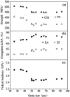
Effect of grain size on (a) yield strength and ultimate tensile strength; (b) on elongation and reduction in area; and (c) on vickers hardness of Ti-29Nb-13Ta-4.6Zr ([144], modified). Reprinted with permission from [144]. Copyright 2002 Springer.
Figure 10.
Figure 10.
Modulus map for Ti-35Nb-7Zr-5Ta based on Nb/Ta ratio vs. Zr content [151]. Reprinted with permission from [151]. Copyright 2006 ASTM International.
Figure 11.
Figure 11.
Stress-controlled fatigue response for Ti-35Nb-7Zr-5Ta with two different oxygen contents [155]. Reprinted with permission from [155]. Copyright 1999 Elsevier.
Figure 12.
Figure 12.
Enhanced osteoblast adhesion on ultrafine-grained (UFG) CP Ti compared to CG CP Ti [147]. Reprinted with permission from [147]. Copyright 2004 Elsevier.
Figure 13.
Figure 13.
Influence of density on mechanical properties of porous Ti-10Mo alloys ([189], modified). Reprinted with permission from [189]. Copyright 2013 Elsevier.
Figure 14.
Figure 14.
Yield strength change of manufactured porous materials with macro porosity fraction [194]. Reprinted with permission from [194]. Copyright 2011 Elsevier.
Figure 15.
Figure 15.
Compressive strength and elastic modulus variation with varying macropore size in porous titanium material with 64% porosity [200]. Reprinted with permission from [200]. Copyright 2011 Elsevier.
Figure 16.
Figure 16.
(a) Mean pore wall thickness values of the porous titanium material produced with different size spacers at various relative densities; (b) Oxygen content of porous titanium material with 25% relative density and varying macropore size [200]. Reprinted with permission from [200]. Copyright 2011 Elsevier.
Figure 17.
Figure 17.
Total porosity behavior as a function of both compacting pressure and sintering temperature [222]. Reprinted with permission from [222]. Copyright 2011 Springer.
Figure 18.
Figure 18.
The schematic fabrication process for porous titanium by space holder method.
Figure 19.
Figure 19.
Scanning electron microscopy (SEM) micrograph of porous titanium with a relative density of 0.30 fabricated using space holder method [48]. Reprinted with permission from [48]. Copyright 2002 Cambridge University Press.
Figure 20.
Figure 20.
Schematic illustration of the fabrication process by using SPS and sodium chloride dissolution: (a) sieving of the titanium and sodium chloride powders; (b) mixing of the titanium and sodium chloride powders; (c) processing by the SPS; (d) dissolution of the sodium chloride in water; (e) obtaining of the porous titanium.
Figure 21.
Figure 21.
SEM micrographs of the porous titanium surfaces with the same porosity of 55% but different pore sizes of 125 (a); 250 (b); 400 (c); and 800 μm (d) [224]. Reprinted with permission from [224]. Copyright 2010 John Wiley and Sons.
Figure 22.
Figure 22.
Schematic diagram showing microwave sintering [270]. Reprinted with permission from [270]. Copyright 2013 Elsevier.
Figure 23.
Figure 23.
Schematic representation of the stages involved in combustion synthesis. The compaction acts as the trigger for initiating an explosion, which synthesizes the mix as it propagates ([232], modified).
Figure 24.
Figure 24.
SEM micrographs of the longitudinal section of the porous Ni-Ti SMA with a banded structure showing channels along the propagating direction of combustion wave [282]. Reprinted with permission from [282]. Copyright 2000 Elsevier.
Figure 25.
Figure 25.
SEM microstructures of porous titanium by using MIM method with sodium chloride, sintered at 1150 °C for 2 h under a vacuum of 1.33 × 10⁻³ Pa: (a) 42.4%; (b) 71.6% [285]. Reprinted with permission from [285].Copyright 2009 Elsevier.
Figure 26.
Figure 26.
Optical microscopy (OM) micrographs of porous NiTi alloys with use of 0 (a) and 8.3 wt% (b) ammonium hydrogen carbonate [288]. Reprinted with permission from [288]. Copyright 2007 Elsevier.
Figure 27.
Figure 27.
Cross sectional images of aligned porous structures for different casting times: (a) 20 h: columnar structure (74%); (b) 24 h: co-existing columnar and lamellar structures (69%); (c) 36 h: most columnar structures have turned into lamellar structures (59%); (d) 48 h: lamellar structure (51%) [299]. Reprinted with permission from [299]. Copyright 2012 Elsevier.
Figure 28.
Figure 28.
Schematic representation of the gel casting process [307]. Reprinted with permission from [307]. Copyright 2012 Elsevier.
Figure 29.
Figure 29.
SEM microstructures of pore morphology and distribution in porous Ti-based alloys processed at 32% solid loading: (a) Ti-17.5Mo alloy; (b) Ti-35Nb alloy, respectively [307]. Reprinted with permission from [307]. Copyright 2012 Elsevier.
Figure 30.
Figure 30.
Processing steps for fabricating porous titanium by slurry foaming [311]. Reprinted with permission from [311]. Copyright 2006 China Academic Journal Electronic Publishing.
Figure 31.
Figure 31.
SEM microstructures of porous titanium fabricated by slurry foaming [311]. Reprinted with permission from [311]. Copyright 2006 China Academic Journal Electronic Publishing.
Figure 32.
Figure 32.
Schematic representation of the plasma spraying process [232]. Reprinted with permission from [232]. Copyright 2006 Elsevier.
Figure 33.
Figure 33.
Entangled metallic wire materials with improved homogeneity [57]. Reprinted with permission from [57]. Copyright 2012 Elsevier.
Figure 34.
Figure 34.
Schematic representation of the three-step replication process ([232], modified).
Figure 35.
Figure 35.
SEM microstructure of reticulated porous Ti-6Al-4V produced by sintering of powders deposited onto a temporary polyurethane scaffold [232]. Reprinted with permission from [232]. Copyright 2006 Elsevier.
Figure 36.
Figure 36.
(a) Illustration of the slurry foaming method; (b) photograph of a multilayer porous titanium sample; (c,d) SEM microstructures of the arrow surface of (b) in (c) low and (d) high magnification [322]. Reprinted with permission from [322]. Copyright 2013 Elsevier.
Figure 37.
Figure 37.
Schematic explaining the additive manufacturing by selective electron beam melting used to generate titanium bodies with a cellular structure [55]. Reprinted with permission from [55].Copyright 2008 Elsevier.
Figure 38.
Figure 38.
(a) Schematic depiction of the laser engineered net shaping (LENS) process; (b) typical porous Ti-6Al-4V samples fabricated using LENS [342]. Reprinted with permission from [342]. Copyright 2010 Elsevier.

References

    1. Park J.B. The Biomedical Engineering Handbook. Vol. 1. CRC Press; Boca Raton, FL, USA: 2000. Biomaterials: Introduction; pp. IV1–IV8.
    1. Temenoff J.S., Mikos A.G. Biomaterials: The Intersection of Biology and Materials Science. Pearson/Prentice Hall; Upper Saddle River, NJ, USA: 2008. pp. 235–258.
    1. Hanawa T. Recent development of new alloys for biomedical use. Mater. Sci. Forum. 2006;512:243–248.
    1. Lütjering G., Williams J.C. Titanium. 2nd ed. Springer; Berlin, Germany: 2003. pp. 20–22.
    1. Long M., Rack H.J. Titanium alloys in total joint replacement—A materials science perspective. Biomaterials. 1998;19:1621–1639. - PubMed