. 2017 Aug;14(2):2268-2280.
doi: 10.3892/ol.2017.6468. Epub 2017 Jun 22.

Diagnosis and surgical treatment of retroperitoneal paraganglioma: A single-institution experience of 34 cases

Affiliations

Diagnosis and surgical treatment of retroperitoneal paraganglioma: A single-institution experience of 34 cases

Xiao-Ke Ji et al. Oncol Lett. 2017 Aug.

Abstract

The present study aimed at identifying the clinical, radiological and pathological characteristics of retroperitoneal paragangliomas, and determining the association between the tumor features and the prognosis of patients following surgery. A total of 34 patients with retroperitoneal paragangliomas, who underwent resection between November 1999 and December 2015, were included in the present retrospective study. The patients' demographics, clinical symptoms and signs, tumor functional status, surgical procedure, intraoperative results, tumor pathology, radiological results, and postoperative survival time were recorded and analyzed. Of the 34 patients, the most common type of presenting symptom was abdominal mass (46%), followed by hypertension (39%) and abdominal pain (32%). Functional tumors occurred in 20 patients (59%). Computed tomography (CT) and magnetic resonance imaging revealed soft-tissue masses, with marked enhancement in the arterial phase, indicative of retroperitoneal paragangliomas. The preoperative CT diagnostic accuracy rate between 2010 and 2015 was markedly improved, compared with that between 1999 and 2009. The tumors were primarily located close to the renal arteries and veins surrounding the abdominal aorta and inferior vena cava. With the exception of one malignant paraganglioma, the majority of paragangliomas were positive for chromogranin A, S-100 protein, vimentin and heat-shock protein 90, and exhibited decreased expression of Ki-67 antigen and insulin-like growth factor 2. All tumors were completely removed by surgery. Distant metastasis, but not tumor size, functional status and local invasion, was markedly associated with survival. The preoperative diagnostic accuracy rate of retroperitoneal paragangliomas may be improved by focusing on the predilection sites and CT characteristics. In addition, immunohistochemical markers were useful to determine tumor malignancy. Complete surgical resection was appropriate for all patients and postoperative survival time was identified to be associated with tumor metastasis.

Keywords: paraganglioma; retroperitoneal tumor; surgical resection; survival time.


Figures

Figure 1.
Figure 1.
Representative radiological images of retroperitoneal paragangliomas. (A) CT image of patient no. 11; (B) CT image of patient no. 11; and (C) CT image of patient no. 11 which demonstrate a round demarcated soft tissue mass with cystic degeneration. The inferior vena cava was depressed by the mass and migrated laterally. The parenchyma of the mass exhibited enhancement, primarily in the arterial phase. (D) CT image of patient no. 10; (E) CT image of patient no. 10; and (F) CT image of patient no. 10, revealing a large oval retroperitoneal mass with cystic degeneration. The parenchyma of the mass exhibited enhancement, and thick tortuous arteries and veins were observed inside the tumor. The juncture point where the tumor vein joined the inferior vena cava was observed. (G) CT image of patient no. 24; (H) CT image of patient no. 24; (I) CT image of patient no. 24; and (J) CT image of patient no. 24, demonstrating a high oval retroperitoneal cystic mass on the left. The parenchyma of the mass exhibited enhancement, and thick tortuous arteries and veins were observed inside the tumor. The tumor arteries originated from the spleen artery, left renal artery, abdominal aortic artery and left internal iliac artery. (K) MRI image of patient no. 20; (L) MRI image of patient no. 20; (M) MRI image of patient no. 20; and (N) MRI image of patient no. 20, demonstrating an oval soft tissue mass posterior to the inferior vena cava. The mass exhibited equal intensities on T1WI and T2WI, and cystic degeneration was observed inside the mass. The mass exhibited an enhancement. The vena cava arched and became thin due to the tumor compression. CT, computed tomography; MRI, magnetic resonance imaging; T1WI, T1-weighted image; T2WI, T2-weighted image.
Figure 2.
Figure 2.
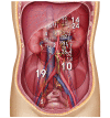
Locations of retroperitoneal paragangliomas in 34 patients in the coronal plane. The number corresponds to the patient number (Table I) and the circle around the number represents the size of the tumor. Retroperitoneal paragangliomas were primarily located close to the renal arteries and veins, surrounding the abdominal aorta and inferior vena cava.
Figure 3.
Figure 3.
Location of retroperitoneal paraganglioma in the horizontal plane. The area of retroperitoneal paragangliomas is highlighted in yellow and all the tumorsidentified in 34 cases were revealed to be within this area.
Figure 4.
Figure 4.
Representative images demonstrating the positive immunostaining of (A) chromogranin-A, (B) S-100 protein, (C) Ki-67, (D) vimentin, (E) heat-shock protein 90 and (F) insulin growth factor 2 from patients with retroperitoneal paragangliomas. Scale bars, 100 µm.
Figure 5.
Figure 5.
Survival rates of patients undergoing resection of retroperitoneal paraganglioma stratified by (A) metastasis, (B) size, (C) functional status, and (D) local invasion.

References

    1. Cunningham SC, Suh HS, Winter JM, Montgomery E, Schulick RD, Cameron JL, Yeo CJ. Retroperitoneal paraganglioma: Single-institution experience and review of the literature. J Gastrointest Surg. 2006;10:1156–1163. doi: 10.1016/j.gassur.2006.05.004. - DOI - PubMed
    1. Lack EE, Cubilla AL, Woodruff JM, Lieberman PH. Extra-adrenal paragangliomas of the retroperitoneum: A clinicopathologic study of 12 tumors. Am J Surg Pathol. 1980;4:109–120. doi: 10.1097/00000478-198004000-00002. - DOI - PubMed
    1. Noda T, Nagano H, Miyamoto A, Wada H, Murakami M, Kobayashi S, Marubashi S, Takeda Y, Dono K, Umeshita K, et al. Successful outcome after resection of liver metastasis arising from an extraadrenal retroperitoneal paraganglioma that appeared 9 years after surgical excision of the primary lesion. Int J Clin Oncol. 2009;14:473–477. doi: 10.1007/s10147-008-0872-1. - DOI - PubMed
    1. Sclafani LM, Woodruff JM, Brennan MF. Extraadrenal retroperitoneal paragangliomas: Natural history and response to treatment. Surgery. 1990;108:1124–1130. - PubMed
    1. Melicow MM. One hundred cases of pheochromocytoma (107 tumors) at the columbia-presbyterian medical center, 1926–1976: A clinicopathological analysis. Cancer. 1977;40:1987–2004. doi: 10.1002/1097-0142(197711)40:5<1987::AID-CNCR2820400502>3.0.CO;2-R. - DOI - PubMed