. 2017 Aug 22;114(34):9170-9175.
doi: 10.1073/pnas.1708558114. Epub 2017 Aug 8.

Antibiotic susceptibility testing in less than 30 min using direct single-cell imaging

Affiliations

Antibiotic susceptibility testing in less than 30 min using direct single-cell imaging

Özden Baltekin et al. Proc Natl Acad Sci U S A. .

Abstract

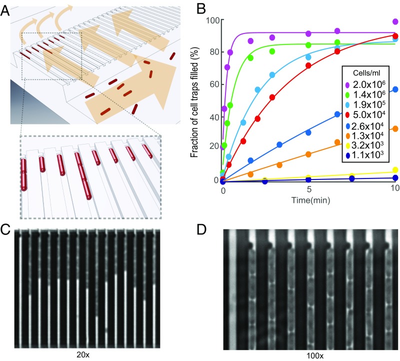
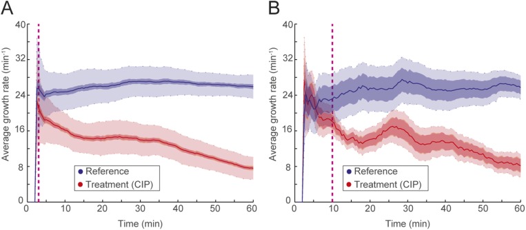
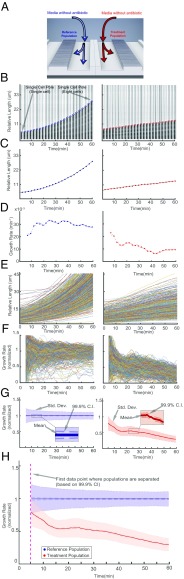
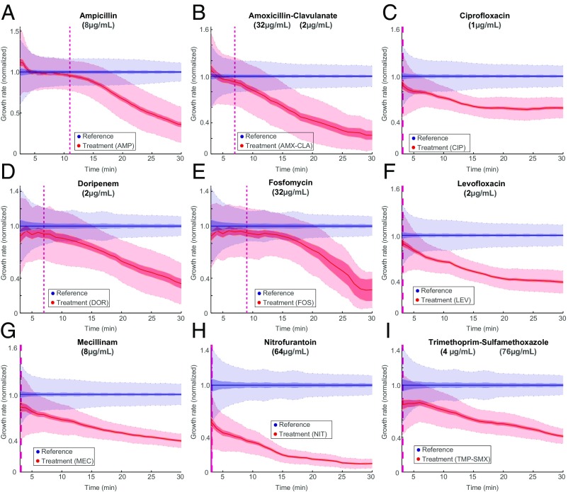
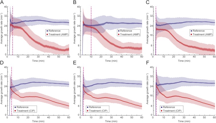
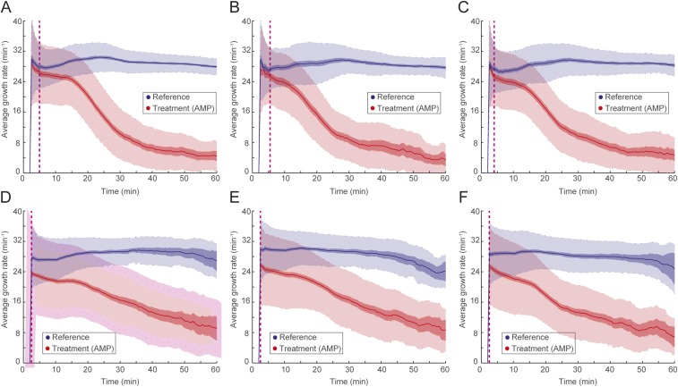
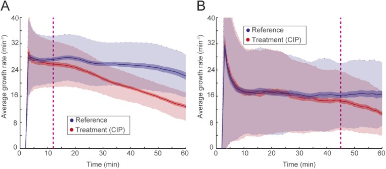
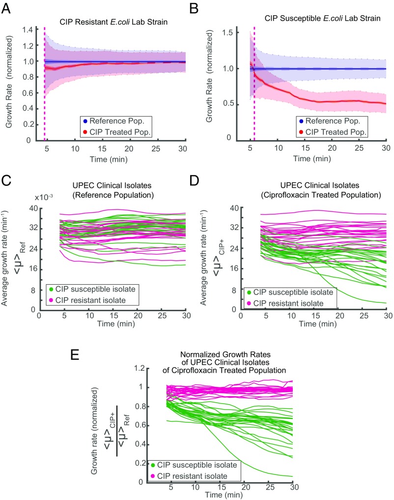
The emergence and spread of antibiotic-resistant bacteria are aggravated by incorrect prescription and use of antibiotics. A core problem is that there is no sufficiently fast diagnostic test to guide correct antibiotic prescription at the point of care. Here, we investigate if it is possible to develop a point-of-care susceptibility test for urinary tract infection, a disease that 100 million women suffer from annually and that exhibits widespread antibiotic resistance. We capture bacterial cells directly from samples with low bacterial counts (104 cfu/mL) using a custom-designed microfluidic chip and monitor their individual growth rates using microscopy. By averaging the growth rate response to an antibiotic over many individual cells, we can push the detection time to the biological response time of the bacteria. We find that it is possible to detect changes in growth rate in response to each of nine antibiotics that are used to treat urinary tract infections in minutes. In a test of 49 clinical uropathogenic Escherichia coli (UPEC) isolates, all were correctly classified as susceptible or resistant to ciprofloxacin in less than 10 min. The total time for antibiotic susceptibility testing, from loading of sample to diagnostic readout, is less than 30 min, which allows the development of a point-of-care test that can guide correct treatment of urinary tract infection.

Keywords: AST; UTI; antibiotic; microfluidic; point of care; resistance.


Conflict of interest statement

Conflict of interest statement: The chip design is being patented (PCT/SE2015/050685). The fast antibiotic susceptibility test is being developed into a product by a company of which Ö.B. and J.E. are shareholders.

Figures

Fig. 1.
Fig. 1.
Design and operation details of the microfluidic chip. (A) Cartoon illustrating the loading of rod-shaped bacterial cells (red) into cell traps. Arrows indicate flow direction during loading. (B) Fraction of cell traps with at least one E. coli cell at different time points. The different markers correspond to different density cell cultures. (C) A phase contrast image of E. coli in the microfluidic device (darker regions) using a 20× objective. (D) A small part of a phase contrast image taken at 100× showing the back end of the cell trap, where the flow restriction region captures the cells during loading.
Fig. S1.
Fig. S1.
2GMM design and features. (A) Drawing of the 2GMM design showing all of the ports with designated port numbers and the orientation of filter regions (yellow) in the ports. Chip number and chip alignment marks are shown in red. (B) Drawing of a port showing the port hole (black) and the filter region. (C) Drawing of a section of a cell trap row.
Fig. S2.
Fig. S2.
2GMM mold scanning electron microscope images with different magnification show some features of 2GMM on the mold, such as (A and F) manifold, (B) two rows of cell traps, (C) cell traps, (D) dot barcodes, and (E and F) filter regions on the ports.
Fig. S3.
Fig. S3.
The fASTest using (A) 951 cells (reference: 409, treatment: 502) and (B) 85 cells (reference: 44, treatment: 41) is compared.
Fig. 2.
Fig. 2.
Detection of growth rate effect of antibiotic. (A) Media with or without antibiotic are supplied to the two different rows of cell traps to test the effect of the antibiotic (CIP; 1 µg/mL) on the treatment population compared with the reference population. (B) A single-cell trap from the reference population (Left) and another single-cell trap from the treatment population (Right) are shown every fifth frame (every 2.5 min). The detected front-most cell pole position is given as a blue or red circle. (C) The corresponding length change through time is shown with the blue or red dots. (D) Growth rates calculated with a sliding window of up to 10 min (starting at 5 min). The (E) length and (F) growth rate are plotted as a function of time for all of the individual traps for the reference (Left) and the treatment population (Right). (G) The descriptive statistics of the normalized growth rate distributions for these populations are shown as a function of time. Colored as blue for the reference population (Left) and red for antibiotic treated population (Right), solid lines show the mean, dark-shaded regions show the 99.9% confidence interval (99.9% CI) of the mean, and the light-shaded region shows the sample SDs. (H) The overlay of the two population’s normalized growth rate distributions. The time of separation of the treatment population from the reference population based on 99.9% CIs occurs before the dashed magenta line, which indicates the first time point when growth rates are estimated.
Fig. 3.
Fig. 3.
Fast detection of response to antibiotic treatment. fASTest experiments testing how fast susceptible E. coli cells respond to (A) ampicillin, (B) AMX-CLA, (C) CIP, (D) DOR, (E) FOS, (F) LEV, (G) MEC, (H) NIT, and (I) TMP-SMX. AMP, ampicillin.
Fig. S4.
Fig. S4.
The fASTest of (A–C) ampicillin and (D–F) CIP for susceptible E. coli (MG1655) biological triplicates. AMP, ampicillin.
Fig. S5.
Fig. S5.
(A–C) The fASTest in standard laboratory medium with three biological replicates of CipR strain using CIP (as reference) and ampicillin and (D–F) the fASTest in urine with three biological replicates of CipR strain to CIP and ampicillin. AMP, ampicillin.
Fig. S6.
Fig. S6.
(A) fASTest for K. pneumoniae and (B) S. saprophyticus for CIP susceptibility detection.
Fig. 4.
Fig. 4.
fASTest for resistant and susceptible strains. Laboratory strains of (A) CIP-resistant and (B) -susceptible E. coli are tested for CIP susceptibility. (C–E) Forty-nine clinical isolates of UPEC are tested with fASTest for CIP susceptibility. (C) Average growth rates for the reference populations. (D) Average growth rates for the treatment populations. Color coding indicates magenta for clinically resistant to CIP and green for clinically susceptible to CIP. (E) Growth rate of treated populations normalized for the growth rate of the respective reference population.
Fig. S7.
Fig. S7.
Image processing steps: (A) region of interest (ROI) detection, (B) cell trap detection, (C) precise ROI detection using the dot barcode, (D) empty trap detection [empty cell trap (I), occupied cell trap (II), template for empty trap averages (III), average empty trap (IV)], and (E) background removal and cell pole detection [occupied cell trap (I), average empty trap (II), enhanced contrast of the occupied trap (III), profile of intensity changes through the contrast enhanced occupied cell trap image (IV)].

References

    1. Kerremans JJ, et al. Rapid identification and antimicrobial susceptibility testing reduce antibiotic use and accelerate pathogen-directed antibiotic use. J Antimicrob Chemother. 2008;61:428–435. - PubMed
    1. Doern GV, Scott DR, Rashad AL. Clinical impact of rapid antimicrobial susceptibility testing of blood culture isolates. Antimicrob Agents Chemother. 1982;21:1023–1024. - PMC - PubMed
    1. van Belkum A, et al. Rapid clinical bacteriology and its future impact. Ann Lab Med. 2013;33:14–27. - PMC - PubMed
    1. Jenkins SG, Schuetz AN. Current concepts in laboratory testing to guide antimicrobial therapy. Mayo Clin Proc. 2012;87:290–308. - PMC - PubMed
    1. Matuschek E, Brown DFJ, Kahlmeter G. Development of the EUCAST disk diffusion antimicrobial susceptibility testing method and its implementation in routine microbiology laboratories. Clin Microbiol Infect. 2014;20:O255–O266. - PubMed

Publication types

MeSH terms