. 2017 Aug 8;7(1):7603.
doi: 10.1038/s41598-017-07855-9.

Regulation of Osteoclast Differentiation by Myosin X

Affiliations

Regulation of Osteoclast Differentiation by Myosin X

Amy Tasca et al. Sci Rep. .

Abstract

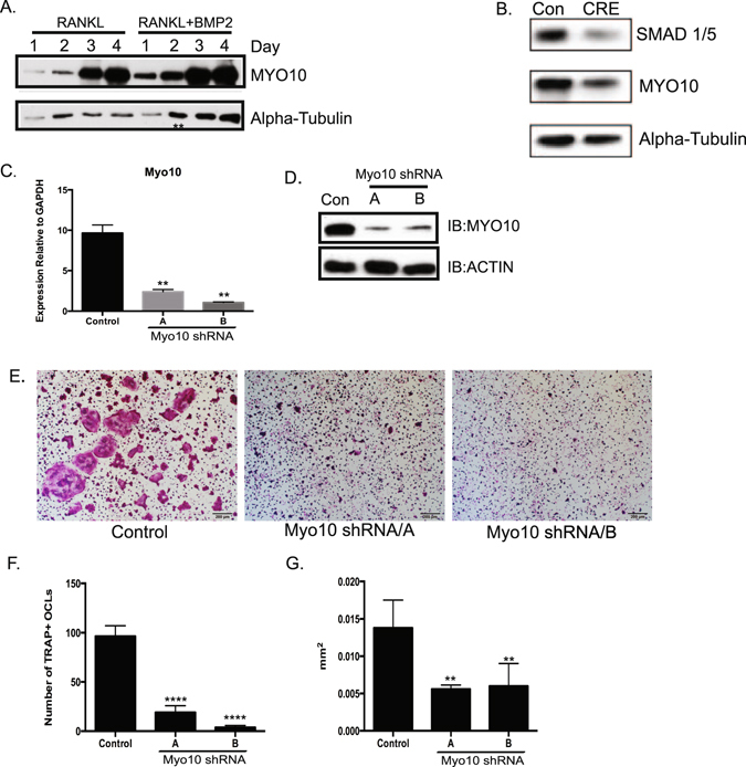
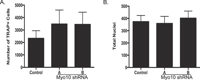
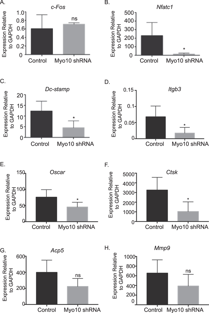
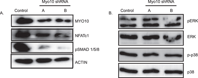
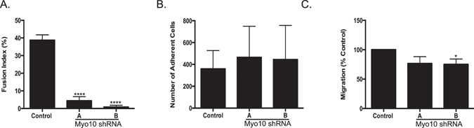
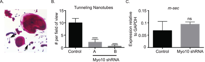
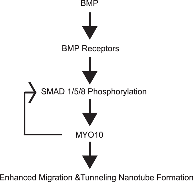
Osteoclasts begin as mononuclear cells that fuse to form multinuclear cells able to resorb bone. The mechanisms that regulate all the steps of osteoclast differentiation are not entirely known. MYO10, an unconventional myosin, has previously been shown in mature osteoclasts to play a role in attachment and podosome positioning. We determined that MYO10 is also expressed early during osteoclast differentiation. Loss of MYO10 expression in osteoclast precursors inhibits the ability of mononuclear osteoclasts to fuse into multinuclear osteoclasts. Expression of Nfatc1, Dc-stamp, Ctsk, and β 3 integrin is reduced in the osteoclasts with reduced MYO10 expression. A slight reduction in the osteoclasts ability to migrate, as well as a reduction in SMAD 1/5/8 phosphorylation are also noted with reduced MYO10 expression. Interestingly we also detected a change in the ability of the osteoclast precursors to form tunneling nanotubes (TNTs), which suggests that MYO10 may regulate the presence of TNTs through its interaction with the cytoskeletal proteins.


Conflict of interest statement

The authors declare that they have no competing interests.

Figures

Figure 1
Figure 1
Myo10 expression is required for osteoclast differentiation. (A) Western blot of osteoclast lysates treated with M-CSF and RANKL (10 ng/mL, left lanes) or M-CSF, RANKL (10 ng/mL) and BMP2 (200 ng/mL, right lanes) for various days. MYO10 and alpha-tubulin expression was analyzed. (B) BMMs were cultured from SMAD1/5 floxed mice and infected with a control or CRE expressing adenovirus. Osteoclasts were treated with M-CSF and RANKL for 3 days. The lysates were analyzed for expression for SMAD1/5 and MYO10 by Western blot. (C) BMMs were cultured from C57Bl/6 mice and infected with lentivirus expressing either a control shRNA or one Myo10 targeting shRNA. Real time RT-PCR was used to measure Myo10 gene expression following 48 hours of infection by lentivirus (D) MYO10 protein levels in shRNA-treated cells were analyzed by western blot (EG) BMMs were differentiated in the presence of M-CSF and RANKL, TRAP stained, imaged and quantified for number and cell area. Only cells with 3 or more nuclei were quantified. Western blots were cropped to show only relevant bands. Experiments were done at least three times and values represent the mean ± SD. **p < 0.01, ****p < 0.0001.
Figure 2
Figure 2
Reduction of Myo10 expression does not affect number of mononuclear osteoclast precursors. BMMs were cultured from C57Bl/6 mice and infected with lentivirus expressing either a control shRNA or one of two Myo10 targeting shRNA. BMMs were differentiated in the presence of M-CSF and RANKL for 2 days. (A) Quantification of day 2 TRAP-stained cells. (B) Quantification of nuclei of day 2 DAPI stained cells. Experiments were done at least three times and values represent the mean ± SD.
Figure 3
Figure 3
Reduction in MYO10 alters key osteoclast gene expression. Real time RT-PCR was used to measure gene expression following 48 hours of infection by lentivirus of (A) c-Fos (B) Nfatc1 (C) Dc-stamp (D) Itgb3 - (E) Oscar (F) Ctsk (G) Acp5 and (H) Mmp9. Experiments were performed at least three times and values represent the mean ± SD. *p < 0.05.
Figure 4
Figure 4
Myo10-deficient osteoclasts undergo less pSMAD1, 5, 8 activation. Western blot of osteoclast extracts that were cultured in the presence of M-CSF and RANKL for 3 days. (A) MYO10, NFATc1, and pSMAD 1/5/8 levels were analyzed by Western blot. Actin was analyzed as a loading control. (B) pERK, ERK, p-p38 and p38 levels were analyzed by Western blot. Western blots were cropped to show only relevant bands. Full length western blots are presented in Supplemental Fig. 4.
Figure 5
Figure 5
Osteoclast fusion and migration are reduced when MYO10 expression is reduced. (A) Fusion index was calculated for day three osteoclasts using the formula fusion index (%) = total number of nuclei within giant cells/total number of nuclei counted × 100. (B) Adhesion of day two primary osteoclasts to vitronectin coated plates. (C) Transwell migration of day two primary osteoclasts. Experiments were performed at least three times and values represent the mean ± SD. *p < 0.05, ****p < 0.0001.
Figure 6
Figure 6
Tunneling nanotube formation is reduced when MYO10 expression is reduced. (A) TRAP image indicating presence of tunneling nanotube. (B) Quantification of tunneling nanotubes at day 2 of culture present as viewed on the 4x objective. (C) RT-PCR quantification of m-sec gene expression of day 2 primary osteoclasts.
Figure 7
Figure 7
The actin cytoskeleton is altered when MYO10 expression is reduced. BMMs were flushed from C57Bl/6 mice and grown on glass coverslips. The cells were infected with lentivirus expressing either a control shRNA or Myo10 targeting shRNA and grown in the presence of M-CSF and RANKL. On day 3 cells were fixed and stained for actin (red) and nuclear DNA (blue) and imaged using confocal microscopy.
Figure 8
Figure 8
Mechanism of MYO10 regulation of osteoclast differentiation. Illustration of a pathway of MYO10 regulation of osteoclast differentiation.

References

    1. Vaananen, H. K. & Laitala-Leinonen, T. Osteoclast lineage and function. Arch Biochem Biophys473, 132–138, doi:S0003-9861(08)00176-8. - PubMed
    1. Boyle WJ, Simonet WS, Lacey DL. Osteoclast differentiation and activation. Nature. 2003;423:337–342. doi: 10.1038/nature01658. - DOI - PubMed
    1. Aguilar PS, et al. Genetic basis of cell-cell fusion mechanisms. Trends Genet. 2013;29:427–437. doi: 10.1016/j.tig.2013.01.011. - DOI - PMC - PubMed
    1. Oursler MJ. Recent advances in understanding the mechanisms of osteoclast precursor fusion. J Cell Biochem. 2010;110:1058–1062. doi: 10.1002/jcb.22640. - DOI - PMC - PubMed
    1. Yagi M, et al. DC-STAMP is essential for cell-cell fusion in osteoclasts and foreign body giant cells. J Exp Med. 2005;202:345–351. doi: 10.1084/jem.20050645. - DOI - PMC - PubMed

Publication types

MeSH terms

LinkOut - more resources