Review
. 2017:128:193-221.

Mast Cells and IgE can Enhance Survival During Innate and Acquired Host Responses to Venoms

Affiliations
Review

Mast Cells and IgE can Enhance Survival During Innate and Acquired Host Responses to Venoms

Stephen J Galli et al. Trans Am Clin Climatol Assoc. 2017.

Abstract

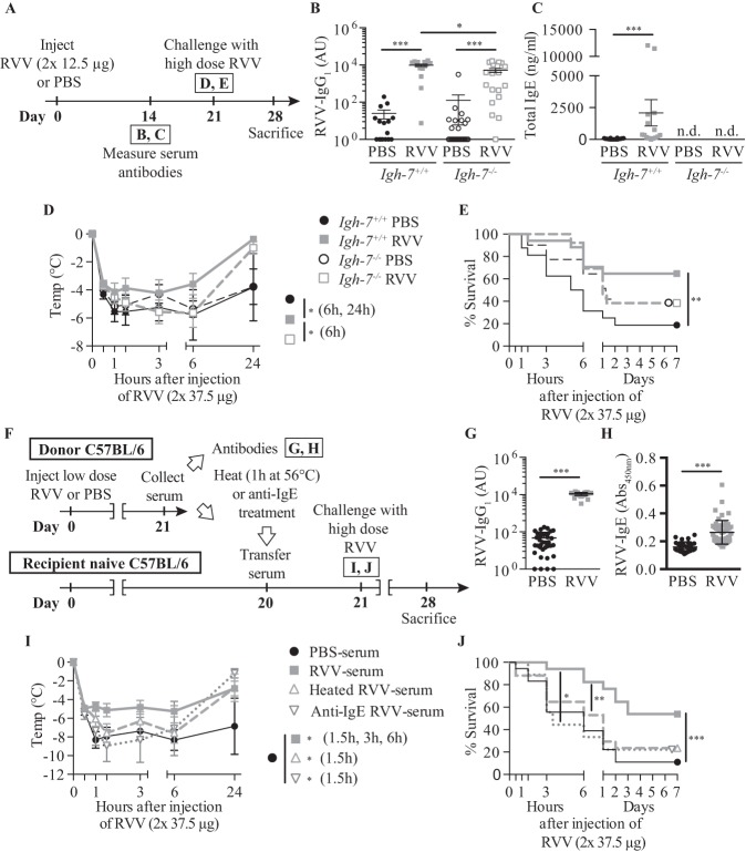
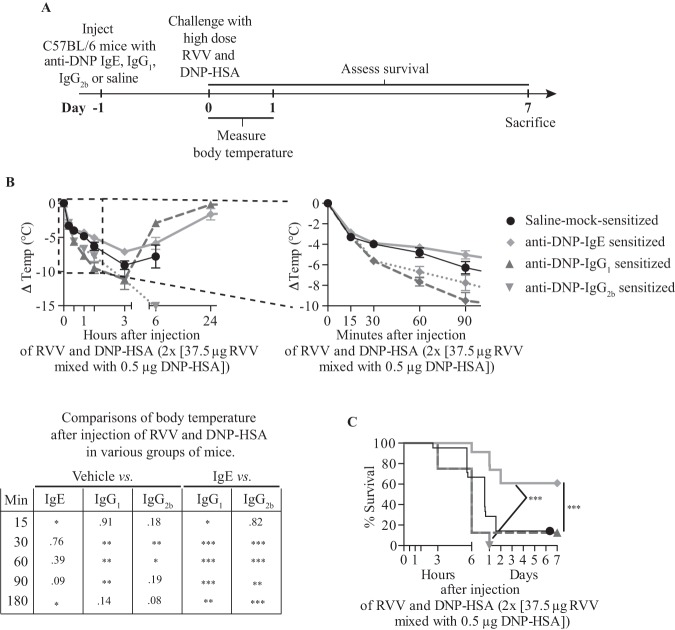
Mast cells and immunoglobulin E (IgE) antibodies are thought to promote health by contributing to host responses to certain parasites, but other beneficial functions have remained obscure. Venoms provoke innate inflammatory responses and pathology reflecting the activities of the contained toxins. Venoms also can induce allergic sensitization and development of venom-specific IgE antibodies, which can predispose some subjects to exhibit anaphylaxis upon subsequent exposure to the relevant venom. We found that innate functions of mast cells, including degradation of venom toxins by mast cell-derived proteases, enhanced survival in mice injected with venoms from the honeybee, two species of scorpion, three species of poisonous snakes, or the Gila monster. We also found that mice injected with sub-lethal amounts of honeybee or Russell's viper venom exhibited enhanced survival after subsequent challenge with potentially lethal amounts of that venom, and that IgE antibodies, FcεRI, and probably mast cells contributed to such acquired resistance.


Conflict of interest statement

Potential Conflicts of Interest: The work reviewed herein was supported by grants to Dr. Galli from the National Institutes of Health (e.g., R37 AI23990, R01 CA072074, R01 AR067145, and U19 AI104209) and the National Science Foundation, and from several other funding sources, including the Department of Pathology at Stanford University. Dr. Starkl was supported by a Max Kade Fellowship of the Max Kade Foundation and the Austrian Academy of Sciences, a Schroedinger Fellowship of the Austrian Science Fund (FWF): J3399-B21, and a Marie Curie fellowship of the European Commission (H2020-MSCA-IF-2014), 655153. Dr. Marichal was supported by a Marie Curie International Outgoing Fellowship for Career Development: European Union’s Seventh Framework Programme (FP7-PEOPLE-2011-IOF), 299954, and a “Charge de recherches” fellowship of the Belgian National Fund for Scientific Research (F.R.S-FNRS).

Figures

Fig. 1
Fig. 1
Making “mast cell knock-in mice.” (1) Mast cells can be generated from bone marrow cells (or other hematopoietic cells, e.g., those in the fetal liver) from wild-type mice or from mutant or transgenic mice with specific genetic alterations of interest (50–52). Alternatively, (2) embryonic stem (ES) cell–derived cultured mast cells (ESCMCs) can be generated from wild-type or genetically altered ES cells (53,54), or (3) various mast cell populations can be transduced in vitro with shRNA to diminish expression of specific genes of interest (15,55). (4) Such bone marrow–, ES cell–, (or fetal liver–) derived cultured mast cells, or shRNA-transduced mast cells, can then be transplanted into mast cell–deficient c-kit mutant mice, such as WBB6F1-KitW/KitW-v mice (52,56) or C57BL/ 6-KitW-sh/KitW-sh mice (57,58), or into C57BL/6-Cpa3-Cre;Mcl-1fl/fl mice (59) (which we informally refer to as “Hello Kitty mice”, which have wild-type c-kit), to produce mast cell knock-in mice. Note: Mouse bone marrow–derived cultured mast cells (BMCMCs) can be injected into genetically mast cell-deficient mice intravenously (i.v.), intraperitoneally (i.p.), or intradermally (i.d.), or into the joints or meninges, etc., but there is a more limited experience with the engraftment of other types of MCs, such as EMCMCs, than with BMCMCs. (5) A suitable interval is then allowed for engraftment and phenotypic “maturation” of the adoptively transferred mast cells (the length of this interval can be varied based on the route of mast cell transfer, the anatomical site of interest, the particular biological response being analyzed, etc.). The importance of mast cell function(s) in biological responses can be analyzed by comparison of the responses in the appropriate wild-type or littermate control mice (6), the corresponding mutant mast cell-deficient mice (7), and selectively mast cell–engrafted mutant mice (mast cell knock-in mice) (8). The contributions of specific mast cell products (surface structures, signaling molecules, secreted products, and so on) to such biological responses can be analyzed by comparing the features of the responses of interest in mast cell knock-in mice engrafted with wild-type mast cells versus mast cells derived from mice or ES cells that lack or express genetically altered forms of such products or that have been transduced with shRNA to silence the specific genes that encode these products. An important part of the analysis of the mast cell knock-in mice used in particular experiments is to assess the numbers and anatomic distribution, and, for certain experiments, aspects of the phenotype, of the adoptively-transferred mast cells, as, depending on the type of in vitro–derived mast cells used, the route of administration, and other factors, these may differ from those of the corresponding native populations of mast cells in the corresponding wild type mice (14,60,61). [This is a modified version of Figure 2 from Metz M, Grimbaldeston MA, Nakae S, et al. Mast cells in the promotion and limitation of chronic inflammation. Immunol Rev 2007;217:304-28 (ref. 60), reprinted with the permission of the publisher, John Wiley and Sons.]
Fig. 2
Fig. 2
Mast cells can diminish Heloderma suspectum venom (H.s.v.)–induced hypothermia and mortality through mast cell protease 4–dependent mechanisms. Changes in rectal temperatures after intradermal injection of H.s.v. (25 mg in 20 ml Dulbecco Modified Eagle Medium [DMEM] solution) into the ear pinnae (one ear pinna of each mouse) of: (A) wild-type (WT) WBB6F1-Kit+/+, mast cell-deficient WBB6F1-KitW/W-v, and WT BMCMCs→KitW/W-v mice (i.e., WBB6F1-KitW/W-v mice which had been engrafted, 6 to 8 weeks before venom challenge, in one ear pinna with 2 million bone marrow–derived cultured mast cells (BMCMCs) derived from WT WBB6F1-Kit+/+ mice) (the death rates of Kit+/+, WT BMCMCs→KitW /W-v, and KitW /W-v mice within 24 hours after H.s.v. injection were 0% [0/21], 7% [1/15, P = 0.42 vs. Kit+/+ mice], and 65% [13/20, P <0.0001 vs. Kit+/+ mice], respectively); (B) WT C57BL/6-Kit+/+, mast cell–deficient C57BL/6-KitW-sh/W-sh, WT BMCMCs→KitW-sh/W-sh, and Mcpt4-/- BMCMCs→KitW-sh/W-sh mice (the death rates of Kit+/+, WT BMCMCs→KitW-sh/W-sh, Mcpt4-/- BMCMCs→KitW-sh/W-sh, and KitW-sh/W-sh mice within 24 hours after H.s.v. injection were 5% [1/19], 11% [2/18, P = 0.48 vs. Kit+/+ mice], 43% [6/14, P = 0.01 vs. Kit+/+ mice], and 50% [10/20, P = 0.006 vs. Kit+/+ mice], respectively); or (C) WT C57BL/6-Kit+/+ mice, C57BL/6-Cpa3Y356L,E378A mice (which have a catalytically inactive CPA3) and C57BL/6-Mcpt4-/- mice (the death rates of Kit+/+, Cpa3Y356L,E378A, and Mcpt4-/- mice within 24 hours after H.s.v. injection were 7% [1/15], 0% [0/14, P = 0.52 vs. Kit+/+ mice], 40% [6/15, P = 0.007 vs. Kit+/+ mice], respectively). Each figure shows data pooled from at least three independent experiments with each group of mice (n = 2-5 mice per group per each individual experiment). **P <0.01, ***P <0.001 versus WT WBB6F1-Kit+/+ or WT C57BL/6-Kit+/+ mice; †P <0.01~0.001 versus each other group (A–C). (D) Extensive degranulation of mast cells (some indicated by closed arrowheads) 1 hour after intradermal injection of H.s.v. (25 mg in 20 ml DMEM), but not vehicle (DMEM) alone (mast cells without evidence of degranulation are indicated by open arrowheads) in WT C57BL/6 mice (Toluidine blue stain; scale bar: 50 µm). (E) Degranulation of mast cells 60 minutes after intradermal injection of H.s.v. (25 mg in 20 ml DMEM) or vehicle (DMEM) alone in WT C57BL/6, Mcpt4-/-, or Cpa3Y356L,E378A mice (injection was into one ear pinna of each mouse). ***P <0.001 versus corresponding vehicle-injected groups; NS = not significant (P >0.05) versus values for WT mice. [This is a reproduction of Figure 1 from Akahoshi M, Song CH, Piliponsky AM, et al. Mast cell chymase reduces the toxicity of Gila monster venom, scorpion venom, and vasoactive intestinal polypeptide in mice. J Clin Invest 2011;121:4180-91 (reference 18), reprinted with the permission of the publisher, the American Society for Clinical Investigation.]
Fig. 3
Fig. 3
Mast cells can enhance innate resistance to high levels of endogenous peptides and structurally similar peptides in reptile venoms. Mast cell cytoplasmic granules contain proteases such as carboxypeptidase A3 (CPA3 [mCPA3 = mouse CPA3]) and mast cell protease 4 (MCP4 [mMCP4 = mouse MCP4]) that, upon secretion by activated mast cells, can degrade certain endogenous peptides, such as endothelin-1 (ET-1) and vasoactive intestinal polypeptide (VIP), respectively, as well as structurally similar peptides contained in the venoms of poisonous reptiles, such as sarafotoxin 6b in the venom of the Israeli mole viper (Atractaspis engaddensis) and helodermin in the venom of the Gila monster (Heloderma suspectum). The ability of mast cells to be activated to degranulate by components of venoms such as these, which can act at the same receptors which recognize the corresponding structurally similar endogenous peptides, permits mast cells to release proteases that can reduce the toxicity of these peptides and which help to enhance the survival of mice injected with the whole venoms of these reptiles, that contain many toxins in addition to sarafotoxin 6b and helodermin. This mechanism may also permit mast cells to restore homeostasis in settings associated with markedly increased levels of the endogenous peptides. [This is a reproduction, in modified form, of Figure 4 from Galli SJ. Rous-Whipple Award Lecture. The mast cell-IgE paradox: from homeostasis to anaphylaxis. Am J Pathol 2016;186:212-24 (reference 42), reprinted with the permission of the publisher, Elsevier, for the American Society for Investigative Pathology.]
Fig. 4
Fig. 4
Evidence that immunoglobulin E (IgE) antibodies contribute to acquired enhanced resistance to the toxicity and lethality of Russell’s viper venom (RVV). (A) Outline of experiments with IgE-deficient (Igh-7-/-) and control (Igh-7+/+) C57BL/6 mice (B-E). (B,C) Serum RVV-specific immunoglobulin G1 (IgG1) (B) and total IgE (C). (D) Body temperature. (E) Survival. (F) Outline of serum transfer experiments in C57BL/6 mice (G–J). (G) Serum RVV-specific IgG1. (H) Serum total IgE. (I) Body temperature. (J) Survival. Data were pooled from three to four experiments (n = 9-25/group). P values: Mann-Whitney test (B, C, G, H), Student t test (D, I) and Mantel-Cox test (E, J). Abbreviation: PBS, phosphate-buffered saline. [This is a reproduction of Figure 3 from Starkl P, Marichal T, Gaudenzio et al. IgE antibodies, FcεRIα and IgE-mediated local anaphylaxis can limit snake venom toxicity. J Allergy Clin Immunol 2016;137:246-57.e11. (reference 93), reprinted with the permission of the publisher, Elsevier.]
Fig. 5
Fig. 5
Immunoglobulin E (IgE)–dependent local mast cell activation induced by activation with a single antigen can enhance resistance to the lethality of Russell’s viper venom (RVV). (A) Experimental outline. (B) Body temperature and (C) Survival of C57BL/6 mice treated with 3 subcutaneous injections of saline alone or containing 50 ng anti-dinitrophenyl (anti-DNP) IgE, IgG1 or IgG2b antibody and challenged 18 hours later with 2 subcutaneous injections, each containing 37.5 µg RVV and 0.5 µg dinitrophenylated human serum albumin (DNP-HSA). Data were pooled from two to five independent experiments (n = 10 to 25/group). P values: Student t test (B); Mantel-Cox test (C). Abbreviation: PBS, phosphate-buffered saline.[This is a reproduction of Figure 5 from Starkl P, Marichal T, Gaudenzio et al. IgE antibodies, FcεRIα and IgE-mediated local anaphylaxis can limit snake venom toxicity. J Allergy Clin Immunol 2016;137:246-57.e11. (reference 93), reprinted with the permission of the publisher, Elsevier.]
None

Similar articles

Cited by

References

    1. Pawankar R, Canonica GW, Holgate ST, et al. Allergic diseases and asthma: a major global health concern. Curr Opin Allergy Clin Immunol. 2012;12:39–41. - PubMed
    1. Paul WE, Zhu J, et al. How are TH2-type immune responses initiated and amplified? Nat Rev Immunol. 2010;10:225–35. - PMC - PubMed
    1. Galli SJ, Tsai M, et al. IgE and mast cells in allergic disease. Nat Med. 2012;18:693–704. - PMC - PubMed
    1. Pulendran B, Artis D, et al. New paradigms in type 2 immunity. Science. 2012;337:431–5. - PMC - PubMed
    1. Kinet JP, et al. The high-affinity IgE receptor (FcεRI): from physiology to pathology. Annu Rev Immunol. 1999;17:931–72. - PubMed

LinkOut - more resources