. 2017 Jul 25:10:230.
doi: 10.3389/fnmol.2017.00230. eCollection 2017.

The Calcineurin-Binding, Activity-Dependent Splice Variant Dynamin1xb Is Highly Enriched in Synapses in Various Regions of the Central Nervous System

Affiliations

The Calcineurin-Binding, Activity-Dependent Splice Variant Dynamin1xb Is Highly Enriched in Synapses in Various Regions of the Central Nervous System

Marie-Lisa Eich et al. Front Mol Neurosci. .

Abstract

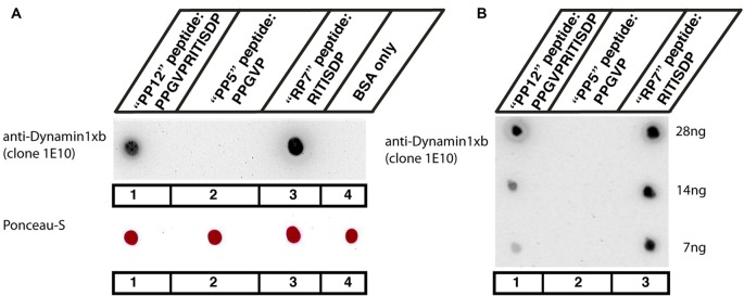
In the present study, we generated and characterized a splice site-specific monoclonal antibody that selectively detects the calcineurin-binding dynamin1 splice variant dynamin1xb. Calcineurin is a Ca2+-regulated phosphatase that enhances dynamin1 activity and is an important Ca2+-sensing mediator of homeostatic synaptic plasticity in neurons. Using this dynamin1xb-specific antibody, we found dynamin1xb highly enriched in synapses of all analyzed brain regions. In photoreceptor ribbon synapses, dynamin1xb was enriched in close vicinity to the synaptic ribbon in a manner indicative of a peri-active zone immunolabeling. Interestingly, in dark-adapted mice we observed an enhanced and selective enrichment of dynamin1xb in both synaptic layers of the retina in comparison to light-adapted mice. This could be due to an illumination-dependent recruitment of dynamin1xb to retinal synapses and/or due to a darkness-induced increase of dynamin1xb biosynthesis. These latter findings indicate that dynamin1xb is part of a versatile and highly adjustable, activity-regulated endocytic synaptic machinery.

Keywords: calcineurin; darkness-induced synaptic recruitment of dynamin1xb; dynamin1xb; retina; splice variant; synapse.


Figures

Figure 1
Figure 1
(A,B) Dot blot analyses of the indicated peptides for their reactivity with the dynamin1xb antibody. Three different dynamin1xb peptides with the indicated amino acid (aa) sequence (“PP12”: PPGVPRITISDP, “PP5”: PPGVP, “RP7”: RITISDP) cross-linked to bovine serum albumin (BSA) were spotted on nitrocellulose membrane. BSA only, i.e., BSA with no peptides cross-linked to it, served as a further control in (A). As expected, the dynamin1xb antibody strongly reacted with the 12mer dynamin1xb peptide (“PP12”) that was used for immunization (spot #1). The antibody also strongly reacted with the dynamin1xb specific carboxyterminal peptide “RP7” that is specific for dynamin1xb (spot #3) but not with the 5mer peptide “PP5” that is common to both dynamin1xa and dynamin1xb (spot #2). The dynamin1xb antibody also did not react with BSA alone (spot #4). Fifty microgram of cross-linked peptide were spotted in (A). Despite the high amount of the peptides spotted in (A), the dynamin 1xb selectively reacts with RP7 (spot #3), the dynamin1xb-specific peptide region but not with “PP5” (spot #2). (B) Similar dot blot analyses as also shown in (A) but with less conjugated peptide spotted to the nitrocellulose membrane. The antibody against dynamin1xb selectively detects the “RP7” peptide of dynamin1xb down to an amount of 7 ng. The sensitivity towards the “RP7” peptide appears to be even higher than the sensitivity towards the “PP12” peptide as judged by the immunostaining intensity of the respective peptide spots with the dynamin1xb antibody.
Figure 2
Figure 2
(A) The indicated mouse tissues were tested for the presence of dynamin1xb with the characterized monoclonal antibody 1E10 by western blot analyses. We observed a single band at the expected running position of ≈100 kDa in neuronal tissues (cerebellum, retina, spinal cord, neocortex, lanes 3–6) but not in extra-neuronal tissues (kidney, intestine; lanes 1,2). In (B) the same tissue extracts as in (A) were loaded and tested subsequently with the dynamin1xb antibody and a monoclonal antibody against actin, that served as a loading control. Similar as in (A), dynamin1xb was only present in the neuronal tissues whereas the actin immunosignal was present in all tissues at ≈43 kDa. (C) The specificity of the dynamin1xb immunolabeling was further corroborated by blocking experiments. The ≈100 kDa band disappeared if the dynamin1xb was pre-absorbed with the dynamin1xb “PP12” peptide (lanes 2,4,6,8) but was unaffected if the antibody was pre-absorbed with an unrelated peptide (lanes 1,3,5,7). Immunodetection of actin at ≈43 kDa served as a loading control.
Figure 3
Figure 3
Semi-thin (0.5 μm-thin) sections of the mouse retina immunolabeled with the monoclonal antibody against dynamin1xb. Very predominantly, the synaptic layers of the retina, the OPL and the IPL were immunolabeled by the dynamin1xb antibody. The synaptic layers were visualized by double-immunolabeling with rabbit polyclonal antibodies against RIBEYE (U2656; Schmitz et al., 2000), a major component of synaptic ribbons. In the synaptic layers, the dynamin1xb signal was discrete and displayed a spot-like distribution at low magnification (A). High-resolution confocal microscopy (B) of the dynamin1xb immunosignals in the OPL where photoreceptor ribbon synapses are located revealed that the dynamin1xb immunolabeling is highly enriched in close vicinity to the synaptic ribbon. The labeling pattern is very similar to the previously observed immunolabeling pattern with a dynamin antibody that did not discriminate between distinct splice variants, e.g., dynamin1xa and dynamin1xb (Wahl et al., 2013). Figure 3 was obtained by confocal microscopy. Abbreviations: ONL, outer nuclear layer; OPL, outer plexiform layer; INL, inner nuclear layer; IPL, inner plexiform layer; GCL, ganglion cell layer. Scale bars: 20 μm (A); 1 μm (B).
Figure 4
Figure 4
Semi-thin (0.5 μm-thin) sections of the mouse retina immunolabeled with the monoclonal dynamin1xb antibody that was pre-absorbed either with a control peptide (A) or with the dynamin1xb peptide “PP12” against which the monoclonal antibody was raised (B). The strong dynamin1xb immunosignal is completely absent if the antibody against dynamin1xb is pre-absorbed with “PP12” whereas the synaptic immunolabel is completely unaffected if a control peptide was used. The RIBEYE immunolabeling was unaffected by both of these treatments. Figure 4 was obtained by confocal microscopy. Abbreviations: ONL, outer nuclear layer; OPL, outer plexiform layer; INL, inner nuclear layer; IPL, inner plexiform layer. Scale bars: 20 μm.
Figure 5
Figure 5
Semi-thin (0.5 μm-thin) sections of the mouse cerebellum double-immunolabeled with the monoclonal dynamin1xb antibody and the indicated other primary antibodies. The other primary antibodies against synaptotagmin1 (A,B), synaptic vesicle protein 2 (SV2; C) and RIM1/2 (D) were applied to label the synapses in order to better relate the dynamin1xb immunosignals to the synaptic regions. We observed a strong dynamin1xb immunosignal in the cerebellar cortex whereas the cerebellar medulla (white matter) that contains predominantly fiber tracts (but no synapses) was not immunolabeled. In the cerebellar cortex, dynamin1xb was highly enriched in the synaptic regions, i.e., the molecular layer (mol) of the cerebellar cortex and the giant synapses in the granule cell layer (arrows) of the cerebellar cortex. No significant dynamin1xb immunosignal was observed in the medulla of the cerebellum that predominantly contains axonal fiber tracts. (A,B,D) was obtained by epifluorescence microscopy; (C) was obtained by confocal microscopy. Abbreviations: mol, molecular layer; Pu, Purkinje cell layer; gr, granule cell layer. Scale bars: 50 μm (A–D).
Figure 6
Figure 6
Semi-thin (0.5 μm-thin) sections of the mouse cerebellum immunolabeled with the monoclonal dynamin1xb antibody that was pre-absorbed either with a control peptide (A) or with the dynamin1xb peptide “PP12” against which the monoclonal antibody was raised (B). The strong dynamin1xb immunosignal in the synaptic regions of the cerebellar cortex was completely absent if the antibody against dynamin1xb was pre-absorbed with “PP12” whereas the synaptic dynamin1xb immunolabel is completely unaffected if a control peptide was used. The SV2 control immunolabeling was completely unaffected by both of these treatments. Figure 6 was obtained by confocal microscopy. Abbreviations: mol, molecular layer; Pu, Purkinje cell layer; gr, granule cell layer. Scale bars: 30 μm.
Figure 7
Figure 7
Semi-thin (0.5 μm-thin) sections of the mouse spinal cord (A–C) and the mouse visual cortex (D) double-immunolabeled with the monoclonal antibody against dynamin1xb and the indicated other primary antibodies. The rabbit polyclonal antibodies against synaptotagmin1 (B,D) and RIM1/2 (A) were applied to label the synapses in the spinal cord. Immunolabeling with rabbit polyclonal antibodies against β-tubulin was used to also visualize the neuronal axons in the white matter of the spinal cord (C). We observed a strong dynamin1xb immunosignal in the gray matter of the spinal cord whereas the white matter that contains many axons (but virtually no synapses) was largely unlabeled by the dynamin1xb antibody. High-resolution confocal analyses revealed the presence of dynamin1xb in synatotagmin1- labeled presynaptic terminals that contact the cell bodies of motor neurons in the gray matter of the spinal cord (B). Similarly, also in the visual cortex (D), we observed a dynamin1xb immunolabeling signal that largely overlapped with synapses as judged by anti-synaptotagmin1 immunolabeling. Arrow in (C) points to an exemplary axon in the white matter of the spinal cord that was immunolabeled with anti-β-tubulin antibodies. (A,C) were obtained by epifluorescence microscopy; (B,D) by confocal microscopy. Abbreviations: n, nucleus of a motor neuron in the anterior horn of the spinal cord; mol, molecular layer; e-gran, external granule cell layer. Scale bars: 50 μm (A,C); 10 μm (B); 30 μm (D).
Figure 8
Figure 8
Semi-thin (0.5 μm-thin) sections of the mouse spinal cord (A,B) or mouse visual cortex (C,D) immunolabeled with the monoclonal antibody against dynamin1xb that was pre-absorbed either with a control peptide (A,C) or with the dynamin1xb peptide “PP12” against which the monoclonal antibody was raised (B,D). The strong dynamin1xb imunosignal in the synaptic layers of the spinal cord and the visual cortex was completely abolished if the antibody against dynamin1xb was pre-absorbed with the “PP12” peptide (B,D) whereas the synaptic immunolabel of dynamin1xb was completely unaffected if a control peptide was used (A,C). Anti-β-tubulin immunolabeling in (A,B) and anti-RIM immunolabeling in (C,D) was completely unaffected by both of these treatments. Figure 8 was obtained by confocal microscopy. Abbreviations: mol, molecular layer; e-gran, external granule cell layer. Scale bars: 10 μm.
Figure 9
Figure 9
Semi-thin (1.5 μm-thin) sections of light- (A) and dark- (B) adapted retinas double-immunolabeled with antibodies against dynamin1xb (A1,3; B1,3) and β-tubulin (A2,3; B2,3). As shown above (Figures 3, 4), the dynamin1xb immunosignal was enriched in the synaptic layers of the retina, the OPL and IPL. In the dark-adapted condition, we observed an enhanced dynamin1xb immunosignal in the synaptic layers of the OPL and IPL, while the β-tubulin immunosignal in the synaptic layers was unchanged. The boxed regions (white boxes) indicate the regions of interest, i.e., OPL and IPL, used for the quantification of immunofluorescence (IF) signals. (A,B) were obtained by confocal microscopy. (C) Quantification of the immunosignals in the OPL and IPL for dynamin1xb and β-tubulin (normalized data). Quantification of IF signals (detemined as integrated density) was done as previously described (Wahl et al., 2016). N = 3 embeddings for light- and dark-adapted retinas; n = 97 images analyzed for both light-and dark-adapted retinas. Error bars are SEM. Abbreviations: A.U, arbitrary units; OPL, outer plexiform layer; INL, inner nuclear layer; IPL, inner plexiform layer; GCL, ganglion cell layer. ***p < 0.001; n.s., non significant. Scale bars: 20 μm.

Similar articles

Cited by

References

    1. Alpadi K., Kulkarni A., Namjoshi S., Srinivasan S., Sippel K. H., Ayscough K., et al. . (2013). Dynamin-SNARE interactions control trans-SNARE formation in intracellular membrane fusion. Nat. Commun. 4:1704. 10.1038/ncomms2724 - DOI - PMC - PubMed
    1. Ames A., III, Parks J. M., Nesbett F. B. (1980). Synthesis and degradation of retinal proteins in darkness and during photoc stimulation. J. Neurochem. 35, 143–148. 10.1111/j.1471-4159.1980.tb12499.x - DOI - PubMed
    1. Anantharam A., Axelrod D., Holz R. W. (2012). Real-time imaging of plasma membrane deformations reveals pre-fusion membrane curvature changes and a role for dynamin in the regulation of fusion pore expansion. J. Neurochem. 122, 661–671. 10.1111/j.1471-4159.2012.07816.x - DOI - PMC - PubMed
    1. Anantharam A., Bittner M. A., Aikman R. L., Stuenkel E. L., Schmid S. L., Axelrod D., et al. . (2011). A new role for the dynamin GTPase in the regulation of the fusion pore expansion. Mol. Biol. Cell 22, 1907–1918. 10.1091/mbc.E11-02-0101 - DOI - PMC - PubMed
    1. Anggono J., Robinson P. J. (2007). Syndapin I and endophilin I bind to overlapping proline-rich regions of dynamin I: role in synaptic vesicle endocytosis. Neurochem 102, 931–943. 10.1111/j.1471-4159.2007.04574.x - DOI - PubMed

LinkOut - more resources