. 2017 Jul 24:11:52.
doi: 10.3389/fnsys.2017.00052. eCollection 2017.

Differences between Dorsal and Ventral Striatum in the Sensitivity of Tonically Active Neurons to Rewarding Events

Affiliations

Differences between Dorsal and Ventral Striatum in the Sensitivity of Tonically Active Neurons to Rewarding Events

Kevin Marche et al. Front Syst Neurosci. .

Abstract

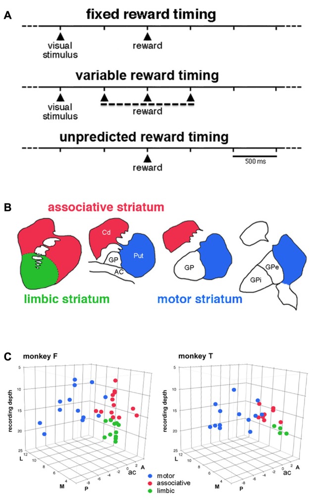
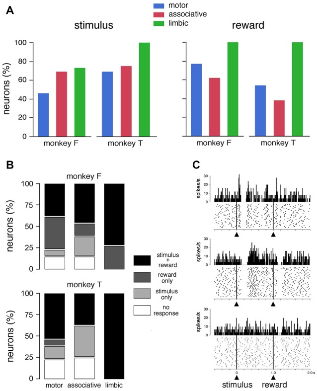
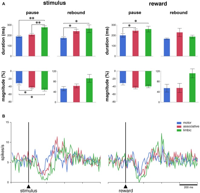
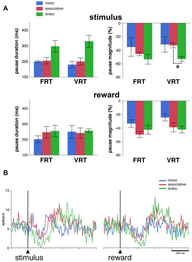
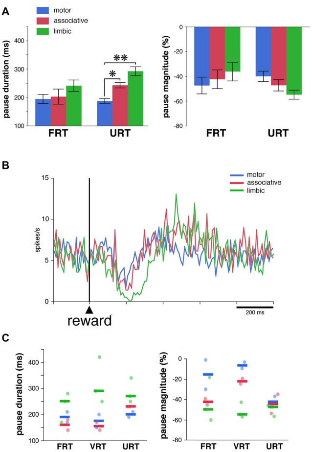
Within the striatum, cholinergic interneurons, electrophysiologically identified as tonically active neurons (TANs), represent a relatively homogeneous group in terms of their functional properties. They display typical pause in tonic firing in response to rewarding events which are of crucial importance for reinforcement learning. These responses are uniformly distributed throughout the dorsal striatum (i.e., motor and associative striatum), but it is unknown, at least in monkeys, whether differences in the modulation of TAN activity exist in the ventral striatum (i.e., limbic striatum), a region specialized for processing of motivational information. To address this issue, we examined the activity of dorsal and ventral TANs in two monkeys trained on a Pavlovian conditioning task in which a visual stimulus preceded the delivery of liquid reward by a fixed time interval. We found that the proportion of TANs responding to the stimulus predictive of reward did not vary significantly across regions (58%-80%), whereas the fraction of TANs responding to reward was higher in the limbic striatum (100%) compared to the motor (65%) and associative striatum (52%). By examining TAN modulation at the level of both the population and the individual neurons, we showed that the duration of pause responses to the stimulus and reward was longer in the ventral than in the dorsal striatal regions. Also, the magnitude of the pause was greater in ventral than dorsal striatum for the stimulus predictive of reward but not for the reward itself. We found similar region-specific differences in pause response duration to the stimulus when the timing of reward was less predictable (fixed replaced by variable time interval). Regional variations in the duration and magnitude of the pause response were transferred from the stimulus to reward when reward was delivered in the absence of any predictive stimulus. It therefore appears that ventral TANs exhibit stronger responses to rewarding stimuli, compared to dorsal TANs. The high proportion of responsive neurons, combined with particular response features, support the notion that the ventral TAN system can be driven by specific synaptic inputs arising from afferent sources distinct from those targeting the dorsal TAN system.

Keywords: basal ganglia; learning; motivation; prediction; primate.


Figures

Figure 1
Figure 1
Timing of events in the testing conditions and approximate functional subdivisions of the striatum. (A) Temporal structure of the sequence of events in the different testing conditions. We used a classical conditioning task in which the presentation of a visual stimulus preceded the delivery of a liquid reward with a constant time interval of 1 s, in the fixed reward timing (FRT) condition, or a pseudorandomly varying interval of 1 ± 0.5 s, in the variable reward timing (VRT) condition. In both conditions, the visual stimulus was extinguished 0.3 s after it came up. In the unpredicted reward timing (URT) condition, the liquid was automatically delivered without preceding stimulus. The three conditions were run in separate blocks of trials and the change in condition was not indicated by any external cues. (B) A drawing illustrating the tripartite subdivision of the primate striatum in the coronal plane, based on previous studies of the topography of the corticostriatal projection in the macaque monkey (Parent and Hazrati, ; Haber and McFarland, 1999). (C) Locations of recorded tonically active neurons (TANs) plotted separately for each monkey. Each color dot represents a single neuron. TANs were recorded between 2 mm anterior (A) and 8 mm posterior (P) to the anterior commissure (ac), over the lateral (L) and medial (M) extent of the striatum measured from the midline (in mm). The recording depth (in mm) was determined from a zero reference point as determined by using microdrive readings. The average depth of penetration of 17 mm was used to divide the precommissural striatum into dorsal and ventral regions.
Figure 2
Figure 2
Responses of TANs to task events in the FRT condition. (A) Proportions of TANs responding to the visual stimulus and reward. Bar plots for each monkey show proportions of responses across the three striatal regions. (B) Relative proportions of TANs showing selective and nonselective responses to stimulus and/or reward. (C) Examples of the responses of TANs to the visual stimulus and/or reward. In each panel, a dot represents a neuronal impulse, and a line of dots represents the neuronal activity recorded during a trial. Histograms and dot displays of TAN activity are aligned on the onset of the stimulus. Vertical calibration on histograms is in spikes per second. Bin width for histograms is 10 ms. The data were taken from monkey T (motor striatum, top panel) and monkey F (ventral striatum, middle and bottom panels).
Figure 3
Figure 3
The responsiveness of TANs to task events in distinct striatal regions. (A) Comparison of durations and magnitudes of the two components of TAN responses (pause and rebound) to the stimulus and reward across striatal regions. Magnitudes of changes are indicated as decreases (pauses) or increases (rebounds) in percentage below baseline activity. Results are pooled for the two monkeys. Values are means ± SEM. **P < 0.01, *P < 0.05. (B) Population average activity of TANs recorded in the three striatal regions. Each curve indicates the mean activity averaged across neurons recorded in a given region for both animals as a function of time from stimulus onset (left) and reward delivery (right). Both the responsive and nonresponsive TANs are included.
Figure 4
Figure 4
Pause responses of TANs in the VRT condition. (A) Effects of a variable stimulus-reward interval on the duration and the magnitude of TAN responses to the stimulus (top) and reward (bottom) in relation to striatal region. Results are pooled for the two monkeys. Values are means ± SEM. *P < 0.05. Same conventions as in Figure 3A. Numbers of neurons responsive to the stimulus and/or reward in the motor, associative and ventral striatum, are as follows: FRT, n = 5–7, 7–8 and 4–6, respectively; VRT, n = 4–6, 8–9, 5–6, respectively. (B) Population average activity across the 27 TANs recorded in the VRT condition. Same conventions as in Figure 3B.
Figure 5
Figure 5
Pause responses of TANs in the URT condition. (A) Same conventions as in Figure 3A. Results are pooled for the two monkeys. Values are means ± SEM. *P < 0.05, **P < 0.01. Numbers of neurons responsive to reward in the motor, associative and ventral striatum, are as follows: FRT, n = 10, 5 and 11, respectively; URT, n = 10, 7, 10, respectively. (B) Population average activity across the 39 TANs recorded in the URT condition. Same conventions as in Figure 3B. (C) Response features of TANs as a function of striatal region in a sample of seven neurons tested in the three conditions. Each dot corresponds to a neuron responsive to reward and thick colored bars represent median values in different striatal regions.

References

    1. Adler A., Katabi S., Finkes I., Prut Y., Bergman H. (2013). Different correlation patterns of cholinergic and GABAergic interneurons with striatal projection neurons. Front. Syst. Neurosci. 7:47. 10.3389/fnsys.2013.00047 - DOI - PMC - PubMed
    1. Aoki S., Liu A. W., Zucca A., Zucca S., Wickens J. R. (2015). Role of striatal cholinergic interneurons in set-shifting in the rat. J. Neurosci. 35, 9424–9431. 10.1523/JNEUROSCI.0490-15.2015 - DOI - PMC - PubMed
    1. Aosaki T., Tsubokawa H., Ishida A., Watanabe K., Graybiel A. M., Kimura M. (1994). Responses of tonically active neurons in the primate’s striatum undergo systematic changes during behavioral sensorimotor conditioning. J. Neurosci. 14, 3969–3984. - PMC - PubMed
    1. Apicella P. (2002). Tonically active neurons in the primate striatum and their role in the processing of information about motivationally relevant events. Eur. J. Neurosci. 16, 2017–2026. 10.1046/j.1460-9568.2002.02262.x - DOI - PubMed
    1. Apicella P. (2007). Leading tonically active neurons of the striatum from reward detection to context recognition. Trends Neurosci. 30, 299–306. 10.1016/j.tins.2007.03.011 - DOI - PubMed

LinkOut - more resources