. 2017 Jul 25:8:540.
doi: 10.3389/fphys.2017.00540. eCollection 2017.

Skin Lesions in Swine with Decompression Sickness: Clinical Appearance and Pathogenesis

Affiliations

Skin Lesions in Swine with Decompression Sickness: Clinical Appearance and Pathogenesis

Long Qing et al. Front Physiol. .

Abstract

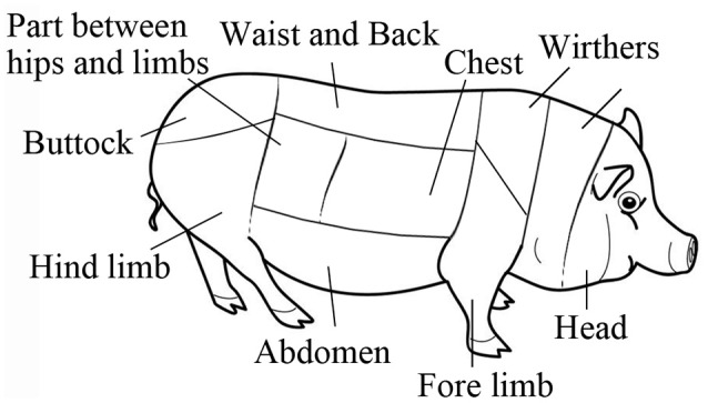
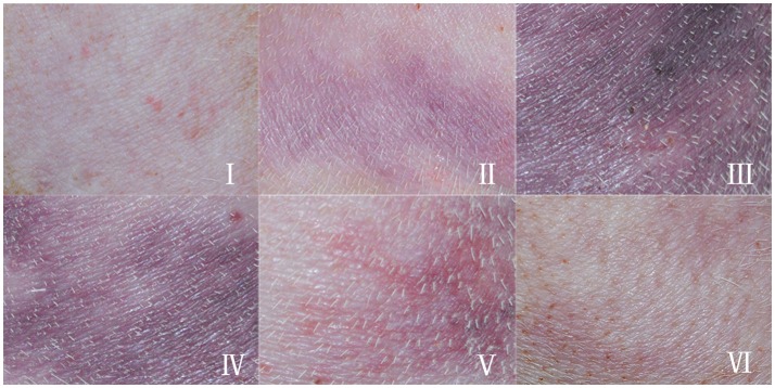
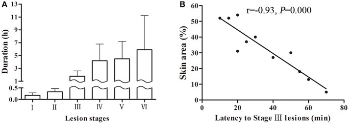
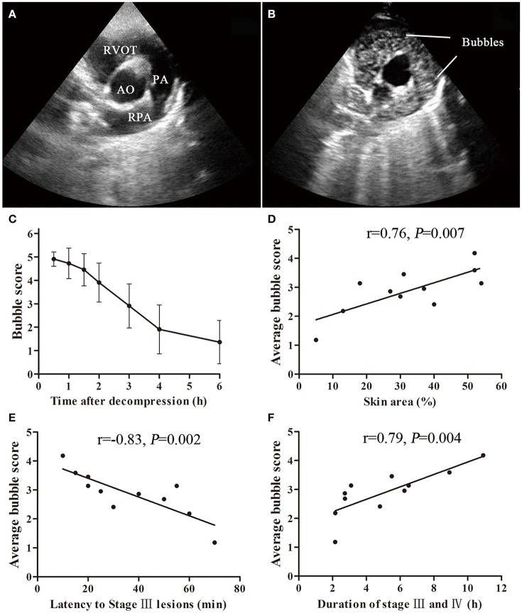
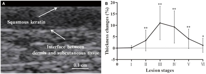
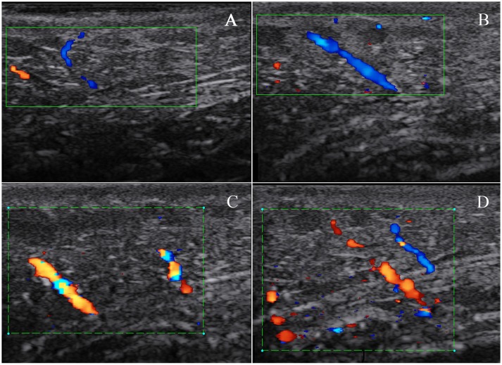
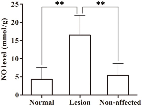
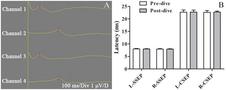
Skin lesions are visual clinical manifestations of decompression sickness (DCS). Comprehensive knowledge of skin lesions would give simple but strong clinical evidence to help diagnose DCS. The aim of this study was to systematically depict skin lesions and explore their pathophysiological basis in a swine DCS model. Thirteen Bama swine underwent simulated diving in a hyperbaric animal chamber with the profile of 40 msw-35 min exposure, followed by decompression in 11 min. After decompression, chronological changes in the appearance of skin lesions, skin ultrasound, temperature, tissue nitric oxide (NO) levels, and histopathology were studied. Meanwhile bubbles and central nervous system (CNS) function were monitored. All animals developed skin lesions and two died abruptly possibly due to cardiopulmonary failure. A staging approach was developed to divide the appearance into six consecutive stages, which could help diagnosing the progress of skin lesions. Bubbles were only seen in right but not left heart chambers. There were strong correlations between bubble load, lesion area, latency to lesion appearance and existence of cutaneous lesions (P = 0.007, P = 0.002, P = 0.004, respectively). Even though local skin temperature did not change significantly, skin thickness increased, NO elevated and histological changes were observed. Increased vessel echo-reflectors in lesion areas were detected ultrasonically. No CNS dysfunction was detected by treadmill walking and evoked potential. The present results suggest skin lesions mainly result from local bubbles and not CNS injuries or arterial bubbles.

Keywords: Bama swine; arterial bubbles; autochthonous bubbles; cutis marmorata; decompression illness; veneous bubbles.


Figures

Figure 1
Figure 1
Body skin map of a swine.
Figure 2
Figure 2
Typical appearance of skin lesions in a swine DCS model. Swine were subjected to a simulated air dive to 40 m for 35 min with a decompression in 11 min, skin appearance were observed and recorded. The hair had been removed prior to the experiment. (I): Erythematous skin, erythematous skin with purple-red discrete macular lesions; (II): Marbling, evolving purple-red marbling; (III): Homogenous purple-red lesion, purple-red homogenous, macular lesion; (IV): Fading lesion, purple-red or blue-black shrinking, and fading macular lesion with reappearance of marbling; (V): Scattered lesion, blue-black scattered, and fading macular lesion; (VI): Remnant, faintly visible blue-black discrete macular lesion.
Figure 3
Figure 3
Skin lesion durations of each stage in a swine DCS model. Eleven swine following a simulated air dive to 40 m for 35 min with 11 min decompression. A total 44 lesions were observed. Durations of each stage are shown (A). The correlation between skin area and latency to Stage III lesions are shown (B).
Figure 4
Figure 4
Bubble formation and correlation with cutaneous lesions in a swine DCS model. Ultrasound bubble detection was performed on 11 swine after a simulated dive to 40 m-35 min with a rapid decompression. An aortic root short axis view was chosen, which shows aorta (AO), right ventricular outflow tract (RVOT), pulmonary artery (PA) and right PA (RPA) (A). Bubbles can be seen in RVOT and PA (B). The bubbles were scored by EB grading scale. Bubble amount decreased slowly during the detecting period from 0.5 to 6 h after decompression (C). Significant correlations between average bubble loads and latency to Stage III lesions, skin area (lesion area/whole body area) or duration of stage III and IV are shown (D–F).
Figure 5
Figure 5
Lesion skin thickness in a swine DCS model. Thickness of skin was determined from the squamous keratin layer to the dermis (A). Forty four lesions were measured. Thickness changes in percentage to the normal are presented as mean ± SD (Compared with normal control *P < 0.05, **P < 0.01) (B).
Figure 6
Figure 6
Subcutaneous blood flow in a swine DCS model. The swine was subjected to a simulated air dive to 40 m for 35 min with an 11 min decompression. Skin blood flow was detected by color Doppler scanning on normal skin in the lateral neck before the dive and repeated on a lesion in the same location. Only two small vessels in subcutaneous tissue were found in normal skin (A). More apparent blood flow signals in the same site were detected on a Stage III lesion, and images in (B–D) show the changes in blood flow signals at different probe angles during detection.
Figure 7
Figure 7
Skin tissue NO levels in a swine DCS model. Tissues were sampled in the lesion and non-affected skin from 11 swine after a simulated air dive to 40 m for 35 min with an 11 min decompression. Normal skin samples were collected pre-dive. NO was detected by ELISA. Values are presented as mean ± SD (**P < 0.01).
Figure 8
Figure 8
Sensory evoked potential in a swine DCS model. Sensory evoked potential (SEP) was tested pre-dive and 6 h post-dive in 11 swine following a simulated air dive to 40 m for 35 min with an 11 min decompression. First lumbar vertebra and head were selected to collect electroneurographic signals from right and left ankles, which were defined as spinal somatosensory evoked potential (SSEP, channels 1 and 3) and cortical somatosensory evoked potential (CSEP, channels 2 and 4) (A). Both sides of SSEP and CSEP were compared pre-dive and post-dive, with no significant changes (B).

References

    1. Bai Y., Mahon R. T., White J. C., Brink P. R., Chon K. H. (2009). Impairment of the autonomic nervous function during decompression sickness in swine. J. Appl. Physiol. 106, 1004–1009. 10.1152/japplphysiol.91246.2008 - DOI - PubMed
    1. Blogg S. L., Gennser M., Møllerløkken A., Brubakk A. O. (2014). Ultrasound detection of vascular decompression bubbles: the influence of new technology and considerations on bubble load. Diving Hyperb. Med. 44, 35–44. - PubMed
    1. Boussuges A., Blanc P., Molenat F., Bergmann E., Sainty J. M. (1996). Haemoconcentration in neurological decompression illness. Int. J. Sports Med. 17, 351–355. 10.1055/s-2007-972859 - DOI - PubMed
    1. Broome J. R., Dick E. J., Jr. (1996). Neurological decompression illness in swine. Aviat. Space Environ. Med. 67, 207–213. - PubMed
    1. Buttolph T. B., Dick E. J., Jr., Toner C. B., Broome J. R., Williams R., Kang Y. H., et al. (1998). Cutaneous lesions in swine after decompression: histopathology and ultrastructure. Undersea Hyperb. Med. 25, 115–121. - PubMed

LinkOut - more resources