

Rapid Maxillary Expansion and Upper Airway Morphology: A Systematic Review on the Role of Cone Beam Computed Tomography
- PMID: 28791305
- PMCID: PMC5534278
- DOI: 10.1155/2017/5460429
Rapid Maxillary Expansion and Upper Airway Morphology: A Systematic Review on the Role of Cone Beam Computed Tomography
Abstract
Objective: This study aimed to investigate the quality of cone beam computed tomography (CBCT) studies evaluating the effects of rapid maxillary expansion on upper airway morphology.
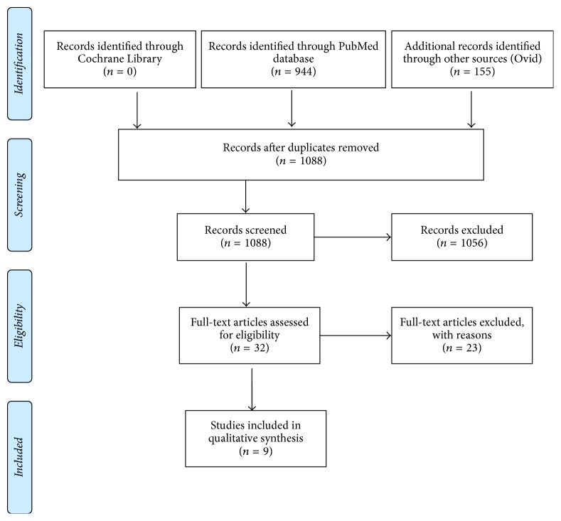
Materials and methods: A database search was conducted using PubMed, Ovid, and Cochrane Library up to December 2016. Studies in which CBCT was adopted to visualize the upper airway before and after rapid maxillary expansion were included. The population target was growing patients. Methodological quality assessment was performed.
Results: The screening process resulted in the exclusion of 1079 references, resulting in only 9 remaining papers that fulfilled the inclusion criteria. No randomized clinical trials were found. The quality scores ranged from 36% to 68% of the maximum achievable, and the mean quality score of the studies was 50%. No good quality studies were detected in our sample.
Conclusions: Inconsistencies in the CBCT protocols utilized were detected between studies. Head posture, tongue position, and segmentation protocols were not consistent. These discrepancies were reflected in the different results obtained in the studies. A valid and consistent protocol with regard to head and tongue positioning, as well as nasal cavity volume segmentation, is required.
Figures
Similar articles
-
Systemic pharmacological treatments for chronic plaque psoriasis: a network meta-analysis.Cochrane Database Syst Rev. 2021 Apr 19;4(4):CD011535. doi: 10.1002/14651858.CD011535.pub4. Cochrane Database Syst Rev. 2021. Update in: Cochrane Database Syst Rev. 2022 May 23;5:CD011535. doi: 10.1002/14651858.CD011535.pub5. PMID: 33871055 Free PMC article. Updated.
-
Effects of rapid maxillary expansion on three-dimensional angular and linear parameters of the Eustachian and auditory tubes in adolescents: A cone-beam computed tomography study.Dent Med Probl. 2025 May-Jun;62(3):435-440. doi: 10.17219/dmp/174822. Dent Med Probl. 2025. PMID: 40569476
-
Orthodontic treatment for posterior crossbites.Cochrane Database Syst Rev. 2001;(1):CD000979. doi: 10.1002/14651858.CD000979. Cochrane Database Syst Rev. 2001. Update in: Cochrane Database Syst Rev. 2014 Aug 08;(8):CD000979. doi: 10.1002/14651858.CD000979.pub2. PMID: 11279699 Updated.
-
Inhaled mannitol for cystic fibrosis.Cochrane Database Syst Rev. 2018 Feb 9;2(2):CD008649. doi: 10.1002/14651858.CD008649.pub3. Cochrane Database Syst Rev. 2018. Update in: Cochrane Database Syst Rev. 2020 May 1;5:CD008649. doi: 10.1002/14651858.CD008649.pub4. PMID: 29424930 Free PMC article. Updated.
-
Intravenous magnesium sulphate and sotalol for prevention of atrial fibrillation after coronary artery bypass surgery: a systematic review and economic evaluation.Health Technol Assess. 2008 Jun;12(28):iii-iv, ix-95. doi: 10.3310/hta12280. Health Technol Assess. 2008. PMID: 18547499
Cited by
-
Rapid Maxillary Expansion and Upper Airway Volume: Systematic Review and Meta-analysis on the Role of Rapid Maxillary Expansion in Mouth Breathing.Int J Clin Pediatr Dent. 2022 Sep-Oct;15(5):617-630. doi: 10.5005/jp-journals-10005-2421. Int J Clin Pediatr Dent. 2022. PMID: 36865716 Free PMC article. Review.
-
Combined orthodontic and surgical open bite correction.Angle Orthod. 2022 Mar 1;92(2):161-172. doi: 10.2319/101921-779.1. Angle Orthod. 2022. PMID: 34986216 Free PMC article.
-
Upper airway changes after rapid maxillary expansion: three-dimensional analyses.BMC Oral Health. 2023 Aug 31;23(1):618. doi: 10.1186/s12903-023-03324-0. BMC Oral Health. 2023. PMID: 37653376 Free PMC article.
-
Novel three-dimensional methods to analyze the morphology of the nasal cavity and pharyngeal airway.Angle Orthod. 2021 May 1;91(3):320-328. doi: 10.2319/070620-610.1. Angle Orthod. 2021. PMID: 33523094 Free PMC article.
-
Effects of rapid maxillary expansion on upper airway volume: A three-dimensional cone-beam computed tomography study.Angle Orthod. 2019 Nov;89(6):917-923. doi: 10.2319/101218-738.1. Epub 2019 Apr 3. Angle Orthod. 2019. PMID: 30942607 Free PMC article.
References
-
- Linder-Aronson S. Respiratory function in relation to facial morphology and the dentition. British Journal of Orthodontics. 1979;6(2):59–71. - PubMed
-
- Saccucci M., Cipriani F., Carderi S., Di Carlo G., D'Attilio M., Rodolfino D., et al. Gender assessment through three-dimensional analysis of maxillary sinuses by means of cone beam computed tomography. European Review for Medical and Pharmacological Sciences. 2015;19(2):185–193. - PubMed
Publication types
MeSH terms
LinkOut - more resources
Full Text Sources
Other Literature Sources
