. 2017 Oct;16(4):4537-4544.
doi: 10.3892/mmr.2017.7177. Epub 2017 Aug 4.

Telmisartan improves vascular remodeling through ameliorating prooxidant and profibrotic mechanisms in hypertension via the involvement of transforming growth factor-β1

Affiliations

Telmisartan improves vascular remodeling through ameliorating prooxidant and profibrotic mechanisms in hypertension via the involvement of transforming growth factor-β1

Peijin Shang et al. Mol Med Rep. 2017 Oct.

Abstract

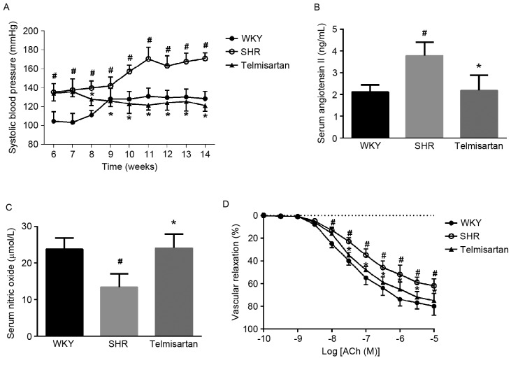
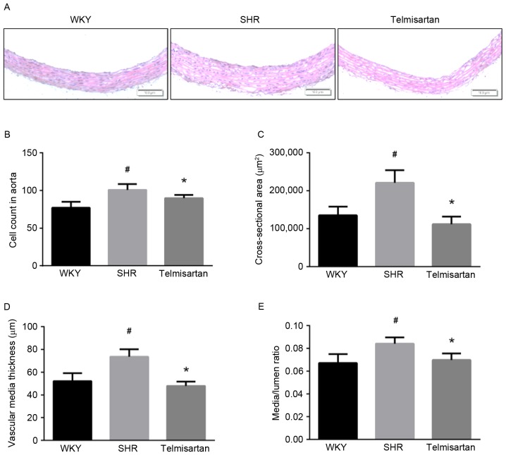
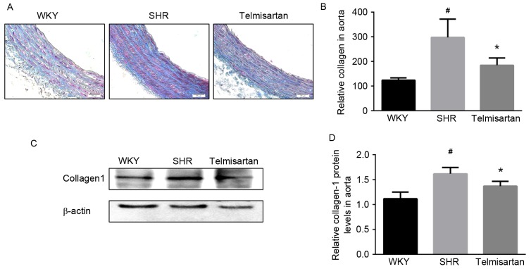
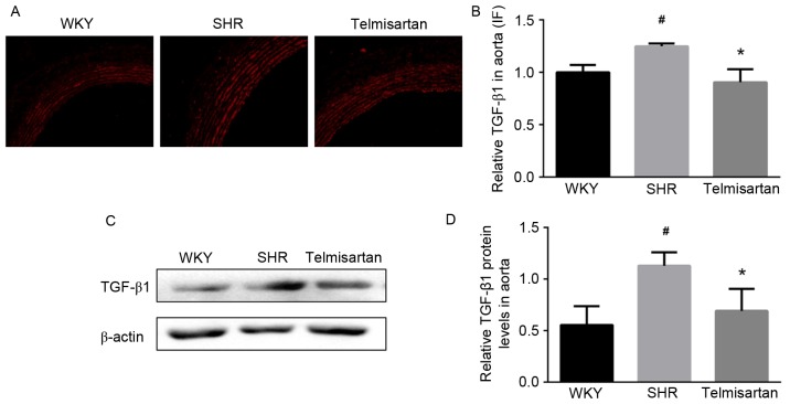
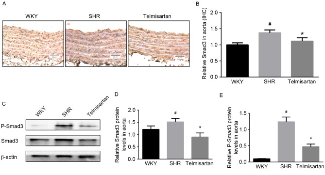
Vascular remodeling is a common complication and pathological basis of hypertension. Transforming growth factor‑β1 (TGF‑β1)/small mothers against decapentaplegic 3 (Smad3) is considered a potential therapeutic target for vascular remodeling in hypertension. The present study aimed to demonstrate the antifibrotic effects of telmisartan and examined the potential mechanisms associated with its prevention of vascular remodeling. Spontaneously hypertensive rats (SHRs) were treated with telmisartan (20 mg/kg), and vascular contractility, reactivity and oxidative stress were preliminarily evaluated. Vascular pathological alterations and collagen deposition were assessed using hematoxylin and eosin, and Masson staining, respectively. The profibrotic factors, TGF‑β1 and Smad3 were evaluated using immunofluorescence and immunohistochemistry. The protein levels of TGF‑β1/Smad3, phosphorylated (p‑)Smad3, collagen‑1 and α-smooth muscle actin in the aorta were assessed using western blot analysis. It was found that telmisartan attenuated chronic vasoconstriction and oxidative stress in the SHRs, and improved vascular reactivity. Telmisartan also restored vascular pathological alterations and decreased collagen deposition. In the vascular wall of the SHRs, telmisartan effectively decreased the protein levels of TGF‑β1/Smad3 and p‑Smad3. Taken together, these findings indicated that telmisartan, with its antioxidant effect, prevented vascular remodeling in hypertension through preventing the TGF‑β1/Smad3 signaling pathway.


Figures

Figure 1.
Figure 1.
Telmisartan decreases SBP and restores vascular reactivity of SHR. (A) Effects on SBP. (B) Angiotensin II levels in serum. (C) Nitric oxide concentration in serum. (D) Vascular reactivity. The results show that telmisartan decreased SBP, attenuated vascular contractility by restoring angiotensin II and nitric oxide in serum, and increased vascular relaxation. Data are presented as the mean ± standard deviation. D. #P<0.05, vs. WKY; *P<0.05, vs. SHR. SBP, systolic blood pressure; WKY, Wistar-Kyoto rats; SHR, spontaneously hypertensive rats.
Figure 2.
Figure 2.
Telmisartan restores vascular morphological alterations. (A) Representative images of Hematoxylin and eosin staining of aortic samples (magnification, ×100). (B) Relative cell count of vascular walls. (C) Vascular cross-sectional area. (D) Vascular wall thickness. (E) Media/lumen ratio. Data are presented as the mean ± standard deviation. #P<0.05, vs. WKY; *P<0.05, vs. SHR. WKY, Wistar-Kyoto rats; SHR, spontaneously hypertensive rats.
Figure 3.
Figure 3.
Telmisartan decreases collagen deposition. (A) Masson staining of aortas (magnification, ×200). (B) Semi-quantitative analysis of collagen deposition. (C) Assessment of protein levels of collagen-1 in aortas using western blot analysis and (D) quantification. Data are presented as the mean ± standard deviation. #P<0.05, vs. WKY; *P<0.05, vs. SHR. WKY, Wistar-Kyoto rats; SHR, spontaneously hypertensive rats.
Figure 4.
Figure 4.
Telmisartan decreases the expression of TGF-β1. (A) Expression of TGF-β1 in the thoracic aorta was assessed using immunofluorescence (magnification, ×200). (B) Semi-quantitative analysis shows that telmisartan significantly decreased the expression of TGF-β1 in the aortas of hypertensive animals. (C and D) Relative protein levels of TGF-β1 in aortas were assessed using western blot analysis. The result showed that telmisartan decreased the expression of TGF-β1. Data are presented as the mean ± standard deviation. #P<0.05, vs. WKY; *P<0.05, vs. SHR. TGF-β1, transforming growth factor-β1; WKY, Wistar-Kyoto rats; SHR, spontaneously hypertensive rats.
Figure 5.
Figure 5.
Telmisartan decreases the levels of Smad3 and p-Smad3. (A) Expression of Smad3 was assessed by immunohistochemistry (magnification, ×200) and (B) optical density was analyzed using Image-Pro Plus 6.0. (C) Western blot analysis revealed that telmisartan effectively decreased the level of (D) Smad3, and inhibited its activation by decreasing the level of (E) p-Smad3. Data are presented as the mean ± standard deviation. #P<0.05, vs. WKY; *P<0.05, vs. SHR. Smad3, small mothers against decapentaplegic 3; p-Smad3, phosphorylated Smad3; WKY, Wistar-Kyoto rats; SHR, spontaneously hypertensive rats.

Similar articles

Cited by

References

    1. Van Bortel LM. What does intima-media thickness tell us? J Hypertens. 2005;23:37–39. doi: 10.1097/00004872-200501000-00009. - DOI - PubMed
    1. Intengan HD, Schiffrin EL. Vascular remodeling in hypertension roles of apoptosis, inflammation, and fibrosis. Hypertension. 2001;38:581–588. doi: 10.1161/hy09t1.096249. - DOI - PubMed
    1. Heusch G, Libby P, Gersh B, Yellon D, Böhm M, Lopaschuk G, Opie L. Cardiovascular remodelling in coronary artery disease and heart failure. Lancet. 2014;383:1933–1943. doi: 10.1016/S0140-6736(14)60107-0. - DOI - PMC - PubMed
    1. Renna NF, de Las Heras N, Miatello RM. Pathophysiology of vascular remodeling in hypertension. Int J Hypertension 2013: Article ID 808353. 2013 doi: 10.1155/2013/808353. - DOI - PMC - PubMed
    1. Montezano AC, Nguyen Dinh Cat A, Rios FJ, Touyz RM. Angiotensin II and Vascular Injury. Curr Hypertens Rep. 2014;16:431. doi: 10.1007/s11906-014-0431-2. - DOI - PubMed

MeSH terms