Review
. 2018 Feb 20;28(6):431-444.
doi: 10.1089/ars.2017.7300. Epub 2017 Sep 8.

Role of Ergothioneine in Microbial Physiology and Pathogenesis

Affiliations
Review

Role of Ergothioneine in Microbial Physiology and Pathogenesis

Bridgette M Cumming et al. Antioxid Redox Signal. .

Abstract

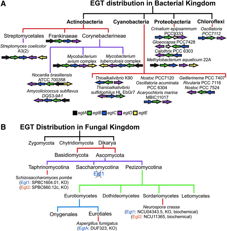
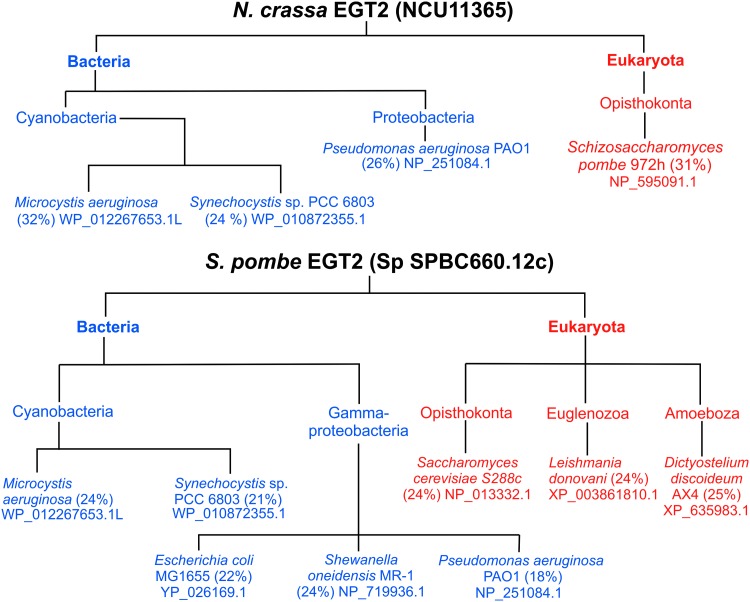
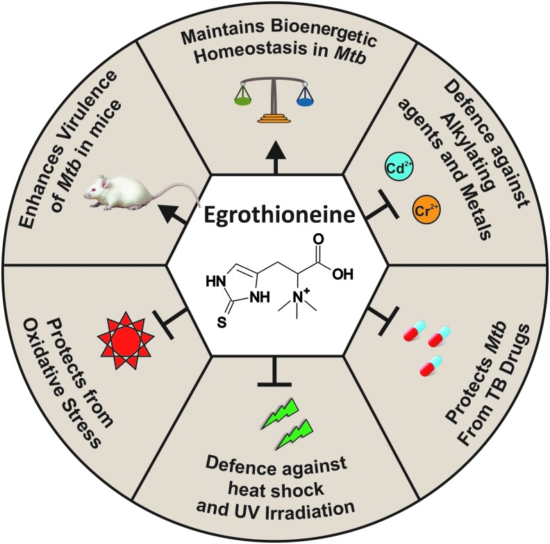
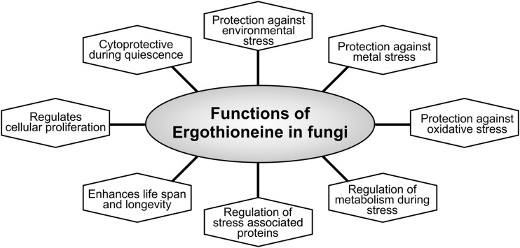
Significance: L-ergothioneine is synthesized in actinomycetes, cyanobacteria, methylobacteria, and some fungi. In contrast to other low-molecular-weight redox buffers, glutathione and mycothiol, ergothioneine is primarily present as a thione rather than a thiol at physiological pH, which makes it resistant to autoxidation. Ergothioneine regulates microbial physiology and enables the survival of microbes under stressful conditions encountered in their natural environments. In particular, ergothioneine enables pathogenic microbes, such as Mycobacterium tuberculosis (Mtb), to withstand hostile environments within the host to establish infection. Recent Advances: Ergothioneine has been reported to maintain bioenergetic homeostasis in Mtb and protect Mtb against oxidative stresses, thereby enhancing the virulence of Mtb in a mouse model. Furthermore, ergothioneine augments the resistance of Mtb to current frontline anti-TB drugs. Recently, an opportunistic fungus, Aspergillus fumigatus, which infects immunocompromised individuals, has been found to produce ergothioneine, which is important in conidial health and germination, and contributes to the fungal resistance against redox stresses.

Critical issues: The molecular mechanisms of the functions of ergothioneine in microbial physiology and pathogenesis are poorly understood. It is currently not known if ergothioneine is used in detoxification or antioxidant enzymatic pathways. As ergothioneine is involved in bioenergetic and redox homeostasis and antibiotic susceptibility of Mtb, it is of utmost importance to advance our understanding of these mechanisms.

Future directions: A clear understanding of the role of ergothioneine in microbes will advance our knowledge of how this thione enhances microbial virulence and resistance to the host's defense mechanisms to avoid complete eradication. Antioxid. Redox Signal. 28, 431-444.

Keywords: ergothioneine; microbes; oxidative stresses; redox homeostasis; thiols.


Figures

<b>FIG. 1.</b>
FIG. 1.
Pie chart indicating the percentage of publications for each naturally occurring low-molecular-weight thiol, excluding glutathione, since 1900 (PubMed search with individual thiol name). Right panel shows the species in which the thiol was first discovered and the year when it was first reported. Ergothioneine is one of the earliest discovered and most extensively studied redox buffers.
<b>FIG. 2.</b>
FIG. 2.
Tautomerism of EGT and chemical structures of oxidation states of EGT. (A) EGT exists as a tautomer between its thiol and thione forms. The thione has resonance structures that are thiolate in character and contribute to weak thiol-like activity of EGT. At physiological pH, EGT exists primarily in the thione form, which prevents it from undergoing autoxidation. (B) ESSE is formed when EGT is exposed to hydrogen peroxide, peroxynitrite, or hypochlorite, but due to its instability at physiological pH, it decomposes disproportionately into EGT and hercynine (C). Servillo et al. (57) proposed two oxidation patterns for EGT: (i) EGT is oxidized via the ESSE pathway and in the presence of excess oxidants, ESSE can be transformed into EGT disulfide S-monoxide (G) and EGT disulfide S-dioxide (H); (ii) EGT is first oxidized into EGT sulfenic acid (E), which is further oxidized into EGT sulfinic acid (F) that is oxidized into EGT sulfonic acid (D); Servillo et al. (57) also proposed a pathway for the decomposition of ESSE at neutral pH (I). EGT, ergothioneine; ESSE, ergothioneine disulfide.
<b>FIG. 3.</b>
FIG. 3.
(A) Five-gene cluster in Mtb H37Rv encoding EGT biosynthetic enzymes; (B) Three protein domains common in egt1 homologs of fungi. (A) An example of the EGT five-gene cluster responsible for EGT biosynthesis that has only been found in Actinobacteria (51, 52). (B) N. crassa Egt1 is a multidomain protein, with an N-terminal SAM-dependent methyltransferase, with 27% identity to M. smegmatis EgtD and C-terminal DinB_2 and FGE-sulfatase domains that have 24% identity to M. smegmatis EgtB (modified from Ref. 9). The domains of NcEgt1 corresponding to Mtb EgtB and EgtD have been indicated. SAM, S-adenosylmethionine.
<b>FIG. 4.</b>
FIG. 4.
Biosynthetic pathways of EGT in (A) mycobacteria and (B) N. crassa and other fungi. (A) In mycobacteria, EGT biosynthesis occurs through five enzymatic steps where L-histidine is methylated into hercynine by methyltransferase (EgtD), followed by formation of a hercynyl γ-glutamylcysteine sulfoxide intermediate after addition of γ-glutamylcysteine by a formylglycine-generating enzyme-like protein (EgtB). Glutamate is removed from this intermediate by a glutamine amidotransferase (EgtC) to form hercynlcysteine sulfoxide that is converted into ergothioneine by a pyridoxal 5-phosphate-dependent β-lyase (EgtE). EgtA is a γ-glutamyl cysteine synthetase. (B) In N. crassa, EGT is synthesized with two enzymes: Egt1 methylates histidine and forms the C-S bond with cysteine to form hercynylcysteine sulfoxide; and Egt2 cleaves the C-S bond to form ergothioneine.
<b>FIG. 5.</b>
FIG. 5.
Distribution of EGT in the (A) bacterial and (B) fungal kingdoms. (A) Distribution of EGT in the bacterial kingdom has been illustrated based on the analysis of the presence or absence of the 5-gene cluster characteristic of Actinobacteria in other bacterial phyla (modified from Ref. 33). (B) Distribution of EGT in the fungal kingdom is illustrated in the main lineages based on orthologs of NcEgt1. Documented evidence for occurrence of EGT is demonstrated in specific species by the annotation of knockout (KO) or biochemical characterization examining the synthesis of EGT in these organisms. No Egt1 homologs were identified in the Saccharomycotina species.
<b>FIG. 6.</b>
FIG. 6.
Homologs of NcEgt2 and SpEgt2 from Blastp searches of model organisms. The amino acid sequence of NcEgt2 and SpEgt2 was used to do a blastp search to discover egt2-like homologs in other microbes that might be responsible for the C-S lyase activity in the final stages of EGT biosynthesis as EgtE, responsible for the C-S lyase activity in mycobacteria, appears to be limited to Actinobacteria. The percentage identity of homologs with the largest coverage of the query sequence is given in brackets and the accession number of the homolog is given below the species name.
<b>FIG. 7.</b>
FIG. 7.
Functions of EGT in bacteria. This figure illustrates functions of ergothioneine in various bacteria, such as maintaining bioenergetic homeostasis in Mycobacterium tuberculosis, modulating susceptibility of M. tuberculosis toward TB drug, protection against diverse oxidative stressors in M. tuberculosis, Methylobacterium aquaticum, and Streptomyces coelicolor, resistance against alkylating agents and metals in M. smegmatis, and defense against heat shock and UV irradiation in M. aquaticum. EGT is essential for survival of M. tuberculosis in macrophages and virulence of M. tuberculosis in mice.
<b>FIG. 8.</b>
FIG. 8.
Functions of EGT in fungi. Schematic overview of the wide physiological functions played by ergothioneine in different fungal species. EGT has been shown to protect conidial germination, thereby enhancing the longevity and overall life span of N. crassa as well as in A. fumigatus. In N. crassa, along with its roles in longevity, EGT is also involved in maintaining cellular redox homeostasis. Similarly, in A. fumigatus, EGT is protective against oxidative stress contributed by free radicals and metal ions such as iron, cobalt, and copper. Furthermore, lack of EGT is also suggested to be responsible for the disruption of iron metabolism in A. fumigatus. In fission yeast, S. pombe, EGT protects the cells during quiescent stages and against environmental stresses, including nitrogen and glucose starvation, and heavy metal stress such as contributed by cadmium (Cd2+).

Similar articles

Cited by

References

    1. Akanmu D, Cecchini R, Aruoma OI, and Halliwell B. The antioxidant action of ergothioneine. Arch Biochem Biophys 288: 10–16, 1991 - PubMed
    1. Alamgir KM, Masuda S, Fujitani Y, Fukuda F, and Tani A. Production of ergothioneine by Methylobacterium species. Front Microbiol 6: 1185, 2015 - PMC - PubMed
    1. Antelmann H. and Helmann JD. Thiol-Based Redox Switches and Gene Regulation. Antioxid Redox Signal 14: 1049–1063, 2011 - PMC - PubMed
    1. Aruoma OI, Whiteman M, England TG, and Halliwell B. Antioxidant action of ergothioneine: assessment of its ability to scavenge peroxynitrite. Biochem Biophys Res Commun 231: 389–391, 1997 - PubMed
    1. Av-Gay Y. and Everett M. The eukaryotic-like Ser/Thr protein kinases of Mycobacterium tuberculosis. Trends Microbiol 8: 238–244, 2000 - PubMed

Publication types

MeSH terms