. 2016 May-Aug;33(3-5):51-63.
doi: 10.1080/09687688.2017.1342969. Epub 2017 Aug 9.

pH-sensitive pHLIP® coated niosomes

Affiliations

pH-sensitive pHLIP® coated niosomes

Mohan C Pereira et al. Mol Membr Biol. 2016 May-Aug.

Abstract

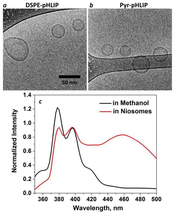
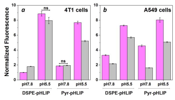
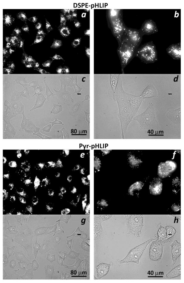
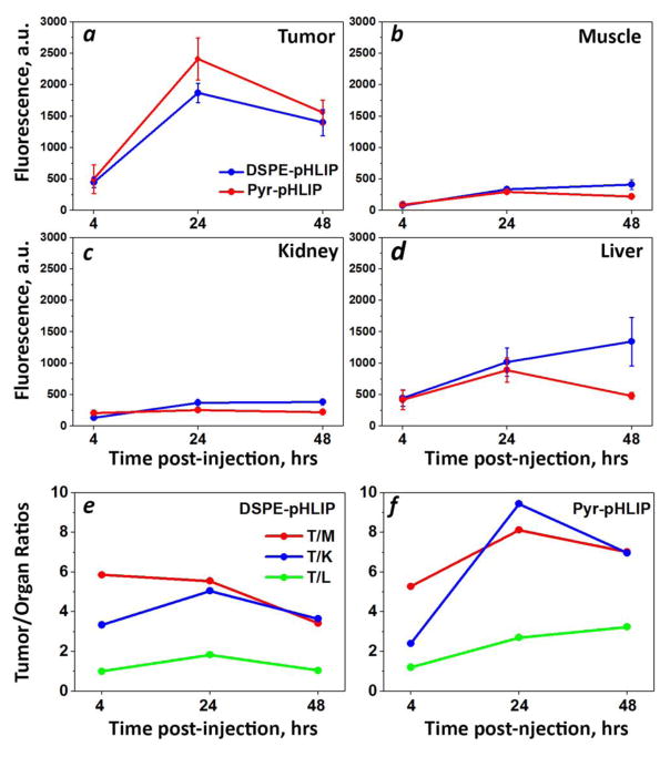
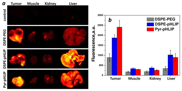
Nanomedicine is becoming very popular over conventional methods due to the ability to tune physico-chemical properties of nano vectors, which are used for encapsulation of therapeutic and diagnostic agents. However, the success of nanomedicine primarily relies on how specifically and efficiently nanocarriers can target pathological sites to minimize undesirable side effects and enhance therapeutic efficacy. Here, we introduce a novel class of targeted nano drug delivery system, which can be used as an effective nano-theranostic for cancer. We formulated pH-sensitive niosomes (80-90 nm in diameter) using nonionic surfactants Span20 (43-45 mol%), cholesterol (50 mol%) and 5 mol% of pH (Low) insertion peptide (pHLIP) conjugated with DSPE lipids (DSPE-pHLIP) or hydrophobic fluorescent dye, pyrene, (Pyr-pHLIP). In coating of niosomes, pHLIP was used as an acidity sensitive targeting moiety. We have demonstrated that pHLIP coated niosomes sense the extracellular acidity of cancerous cells. Intravenous injection of fluorescently labeled (R18) pHLIP-coated niosomes into mice bearing tumors showed significant accumulation in tumors with minimal targeting of kidney, liver and muscles. Tumor-targeting niosomes coated with pHLIP exhibited 2-3 times higher tumor uptake compared to the non-targeted niosomes coated with PEG polymer. Long circulation time and uniform bio-distribution throughout the entire tumor make pHLIP-coated niosomes to be an attractive novel delivery system.

Keywords: Drug delivery; fluorescence imaging; targeting tumor acidity.


Conflict of interest statement

Declaration of Interest

OA Andreev and YK Reshetnyak have founded and have a financial interest in a company, pHLIP, Inc., with the aim of bringing pHLIP technology to the clinic. The company has had no involvement in funding the studies reported here.

Figures

Figure 1
Figure 1
Cryogenic TEM image of the a) DSPE-pHLIP (5 mol%) and b) Pyr-pHLIP (5 mol%) coated Span20 (45 mol%) and cholesterol (50 mol%) niosomes. The images are obtained at 25,000x magnification. c) The fluorescence spectra of Pyr-pHLIP measured in methanol and Pyr-pHLIP incorporated in niosomes.
Figure 2
Figure 2
Normalized uptake of DSPE-pHLIP (5 mol%) and Pyr-pHLIP (5 mol%) coated Span20 (43 mol%) and cholesterol (50 mol%) niosomes containing 2 mol% of fluorescent R18 by 4T1 mammary (a) and A549 lung (b) cancer cells at pH 7.8 and pH 5.5 before (magenta columns) and after (gray columns) treatment with Trypan blue. The fluorescent signals were normalized by the rhodamine fluorescence intensity of 4T1 cells at pH 7.8 treated with DSPE-pHLIP before addition of Trypan blue. Statistically significant differences were determined by two-tailed unpaired Student’s t-test, only statistically non-significant differences are indicated (ns means p-level > 0.05), all other differences in cellular uptake calculated at different pHs, as well as before and after Trypan Blue addition are statistically significant (p-level is less than 0.0001 in each case). The distribution of fluorescent signal in cells is presented in Figure S4.
Figure 3
Figure 3
Uptake and cellular distribution of a–d) DSPE-pHLIP (5 mol%) and e–h) Pyr-pHLIP (5 mol%) coated Span20 (43 mol%) and cholesterol (50 mol%) niosomes containing 2 mol% of fluorescent R18 by A549 lung cancer cells. Cells were treated with fluorescence niosomes at pH 6.4 for 1 hour, followed by washing, seeding cells in glass bottom collagen coated cell dishes and imaging at next day. Fluorescence (a, b, e, f) and phase contrast (c, d, g, h) images were obtained using 20x (a, c, e, g) and 40x (b, d, f, h) magnification objective lenses.
Figure 4
Figure 4
Time-dependent distribution of DSPE-pHLIP and Pyr-pHLIP (5 mol%), Span20 (43 mol%), cholesterol (50 mol%) and R18 (2 mol%) niosomes in a) tumor, b) muscle, c) kidney and d) liver at 4, 24 and 48 hours after single I.V. administration of the constructs. Tumor/Muscle (T/M), Tumor/Kidney (T/K) and Tumor/Liver (T/L) ratio calculated for DSPE-pHLIP (e) and Pyr-pHLIP (f) niasomes are shown. The values of mean surface fluorescence intensity of R18 in tissue and organs are presented in Table S1.
Figure 5
Figure 5
a) The representative rhodamine fluorescence images and b) mean surface fluorescence of tumor (cut in half), muscle, kidney (cut in half) and liver obtained by ex vivo imaging after collection of organs and tissues 24 hours after I.V. administration of pHLIP and PEG coated niosomes are shown (the autofluorescence signal is subtracted). The color coded fluorescent images shown on panel a are obtained at the same settings of the imaging instrument, the same exposure time (15 sec), and all of them were processed exactly the same way. Control means organs collected from the mouse with no injection of fluorescent niosomes, and it represents level of auto fluorescence signals in tissue. The values of mean surface fluorescence intensity of R18 in tissue and organs are presented in Table S2.
Figure 6
Figure 6
Distribution of R18 fluorescent signal in tumor sections obtained at 24 hours after I.V. injection of a–d) DSPE-pHLIP (5 mol%) and e–h) Pyr-pHLIP (5 mol%) coated Span20 (43 mol%), cholesterol (50 mol%) and R18 (2 mol%) niosomes. The rhodamine fluorescence (a, e), cell nucleus stained with DAPI (b, f), the overlay of rhodamine and DAPI fluorescence (c, g), and adjacent HE stained sections from the same tumor (d, h) are shown.

Similar articles

Cited by

References

    1. AMMAR H, GHORAB M, EL-NAHHAS S, HIGAZY I. Proniosomes as a carrier system for transdermal delivery of tenoxicam. International journal of pharmaceutics. 2011;405:142–152. - PubMed
    1. ANDERSON M, MOSHNIKOVA A, ENGELMAN DM, RESHETNYAK YK, ANDREEV OA. Probe for the measurement of cell surface pH in vivo and ex vivo. Proc Natl Acad Sci U S A. 2016;113:8177–81. - PMC - PubMed
    1. ANDREEV OA, ENGELMAN DM, RESHETNYAK YK. Targeting acidic diseased tissue: New technology based on use of the pH (Low) Insertion Peptide (pHLIP) Chimica oggi. 2009;27:34. - PMC - PubMed
    1. ANDREEV OA, ENGELMAN DM, RESHETNYAK YK. pH-sensitive membrane peptides (pHLIPs) as a novel class of delivery agents. Molecular membrane biology. 2010;27:341–352. - PMC - PubMed
    1. ARACHCHIGE MC, RESHETNYAK YK, ANDREEV OA. Advanced targeted nanomedicine. Journal of biotechnology. 2015;202:88–97. - PMC - PubMed