. 2017 Oct 3;13(10):1697-1708.
doi: 10.1080/15548627.2017.1356549. Epub 2017 Aug 9.

SQSTM1/p62-mediated autophagy compensates for loss of proteasome polyubiquitin recruiting capacity

Affiliations

SQSTM1/p62-mediated autophagy compensates for loss of proteasome polyubiquitin recruiting capacity

Alik Demishtein et al. Autophagy. .

Abstract

Protein homeostasis in eukaryotic cells is regulated by 2 highly conserved degradative pathways, the ubiquitin-proteasome system (UPS) and macroautophagy/autophagy. Recent studies revealed a coordinated and complementary crosstalk between these systems that becomes critical under proteostatic stress. Under physiological conditions, however, the molecular crosstalk between these 2 pathways is still far from clear. Here we describe a cellular model of proteasomal substrate accumulation due to the combined knockdown of PSMD4/S5a and ADRM1, the 2 proteasomal ubiquitin receptors. This model reveals a compensatory autophagic pathway, mediated by a SQSTM1/p62-dependent clearance of accumulated polyubiquitinated proteins. In addition to mediating the sequestration of ubiquitinated cargos into phagophores, the precursors to autophagosomes, SQSTM1 is also important for polyubiquitinated aggregate formation upon proteasomal inhibition. Finally, we demonstrate that the concomitant stabilization of steady-state levels of ATF4, a rapidly degraded transcription factor, mediates SQSTM1 upregulation. These findings provide new insight into the molecular mechanisms by which selective autophagy is regulated in response to proteasomal overflow.

Keywords: ADRM1/Rpn13; SQSTM1/p62; autophagy; proteasome; ubiquitin receptor PSMD4/Rpn10.


Figures

Figure 1.
Figure 1.
PSMD4 and/or ADRM1 knockdown increase accumulation of polyubiquitinated cargoes with autophagic markers. (A) HeLa cells stably expressing GFP-LC3B were transfected with either nontargeting siRNA (Scr.) or with PSMD4 and ADRM1 siRNAs separately or together using DharmaFect reagent. After 72 h cells were fixed, immunostained with anti-SQSTM1 and anti-Ub antibodies, and analyzed by confocal microscopy. Scale bar: 10 μm. (B) Immunoprecipitation analysis of endogenous SQSTM1 using anti-SQSTM1. The following antibodies were employed for results visualization by western blot analysis: anti-SQSTM1, anti-Ub and anti-Actin. HeLa cells were transfected with either non-targeting siRNA (Scr.) or mixed PSMD4 and ADRM1 siRNAs using DharmaFect reagent for 72 h. Then total cellular extracts were obtained with NP-40 buffer and used for the immunoprecipitation.
Figure 2.
Figure 2.
Autophagy is involved in the clearance of ubiquitin-labeled proteins accumulated upon knockdown of PSMD4 and/or ADRM1, in an MTORC1-independent manner. Estimation of autophagic flux by (A) western blot analysis and (B) confocal microscopy. HeLa cells were transfected with either nontargeting siRNA (Scr.) or PSMD4 and ADRM1 siRNAs separately or together using DharmaFect reagent for 72 h. To inhibit lysosomal function, 0.1 µM BafA was added for the last 10 h of the experiment. For western blot analyses cells were lysed with NP-40 buffer, subjected to SDS-PAGE, transferred to nitrocellulose membrane and visualized with anti-SQSTM1, anti-LC3, anti-PSMD4, anti-ADRM1 and anti-Actin antibodies. For confocal microscopy cells were fixed and immunostained with anti-SQSTM1 and anti-LC3 antibodies. The confocal figures are representative images chosen from the analysis of more than 120 cells in 3 independent experiments. The autophagic flux of SQSTM1 and LC3-II in panel (A) was quantified using Imagequant™ software. The results are the mean ± STDEV of at least 3 independent experiments. *p < 0.05 for the difference between Scr. and the PSMD4 and ADRM1 siRNA, determined by ANOVA with Dunnet Multiple Comparison Test as post-test. Scale bar: 10 μm. (C) Total RNA of HeLa cells, transfected with non-targeting siRNA (Scr.) or a mix of S5a/ADRM1 siRNAs, was extracted and subjected to quantitative RT-PCR analysis using the indicated primers. The level of the mRNA was normalized to level of HPRT. The results are the mean ± STDEV of at least three independent experiments. *p < 0.05 for the difference between Scr (represented as dashed line at 1) and the S5a/ADRM1 siRNA, determined by ANOVA with Dunnet Multiple Comparison Test as post-test. (D) HeLa cells were transfected with a mix of PSMD4 and ADRM1 siRNAs using DharmaFect reagent for 72 h. The cells were transfected with a plasmid encoding GFP-ATG4C74A 24 h after the siRNA transfection, for an additional 48 h. To inhibit lysosomal function 0.1 µM BafA was added for the last 10 h of the experiment. The cells were fixed, immunostained with anti-SQSTM1 and anti-Ub antibodies, and analyzed by confocal microscopy. Scale bar: 10 μm. (E) Activity of MTOR was estimated by HeLa cells transfected with nontargeting siRNA (Scr.) or mixed PSMD4 and ADRM1 siRNAs using DharmaFect reagent for 72 h. As a control for MTORC1 inactivation HeLa cells were incubated for 4 h in EBSS starvation medium. The cells were lysed with NP-40 buffer supplemented with phosphatase inhibitors and analyzed by western blot with the indicated antibodies. The level of p-ULK1 (S757) was quantified using Imagequant™ software. The results are the mean ± STDEV of at least 3 independent experiments. *P < 0.05 for the difference between Scr. (represented as dashed line at 1), si-PSMD4 and si-ADRM1, and starvation-treated cells was determined by ANOVA with Dunnet Multiple Comparison Test as post-test. *P < 0.01 for the difference between si-PSMD4 and si-ADRM1, and starvation-treated cells was determined by paired t-test. A.U., arbitrary units.
Figure 3.
Figure 3.
Recruitment of polyubiquitinated substrates into phagophores is mediated by SQSTM1, but not NBR1. (A) HeLa cells were transfected with either nontargeting siRNA (Scr.), SQSTM1 siRNA, a mix of PSMD4 and ADRM1 or a mix of PSMD4, ADRM1 and SQSTMQ or NBR1 siRNAs using DharmaFect reagent for 72 h. After 24 h the indicated cells were transfected again with control or SQSTM1 siRNA. Then cells were fixed, immunostained with anti-SQSTM1 and anti-Ub antibodies, and analyzed by confocal microscopy. Scale bar: 10 µm. (B) HeLa cells were transfected with nontargeting siRNA (Scr.), a mix of PSMD4 and ADRM1 or a mix of PSMD4, ADRM1 and NBR1 or SQSTMQ siRNAs using DharmaFect reagent for 72 h. To inhibit lysosomal function, 0.1 µM BafA was added for the last 10 h of the experiment. Next, cells were fixed, immunostained with anti-SQSTM1 and anti-Ub antibodies, and analyzed by confocal microscopy. Scale bar: 10 µm. (C) Total RNA of HeLa cells, transfected with nontargeting siRNA (Scr.) or a mix of PSMD4 and ADRM1 siRNAs, was extracted and subjected to quantitative RT-PCR analysis using SQSTM1, NBR1 and HPRT1 primers. The level of the mRNA was normalized to the level of HPRT1. The results are the mean ± STDEV of at least 3 independent experiments. *p < 0.05 for the difference between Scr (represented as a dashed line at 1) and the PSMD4 and ADRM1 siRNA, determined by ANOVA with Dunnet Multiple Comparison Test as post-test.
Figure 4.
Figure 4.
Knockdown of SQSTM1 protects against aggregate formation after proteasomal inhibition by Velcade. (A) HeLa cells were either transfected with nontargeting siRNA (Scr.) or a mix of PSMD4 and ADRM1 siRNAs using DharmaFect reagent for 72 h. To inhibit proteasomal function, 2.5 µM velcade was added for 15 h to untransfected cells. Then cells were lysed with NP-40 extraction buffer and the extracts were centrifuged for 20 min at 20,000 g. The supernatant (left panel) and pellet (right panel) fractions were analyzed by western blot using anti poly-Ub, anti-SQSTM1, anti-TP53 and anti-Actin antibodies. (B) HeLa cells were either transfected with nontargeting siRNA (Scr.) or SQSTM1 siRNAs using DharmaFect reagent for 72 h. After a 24-h interval the cells were transfected once again with SQSTM1 siRNA. To inhibit proteasomal function, 2.5 µM Velcade was added for the last 15 h of the experiment. Then cells were lysed with NP-40 extraction buffer and the extracts centrifuged for 20 min at 20,000 g. The supernatant (left panel) and pellet (right panel) fractions were analyzed by western blot using anti poly-Ub, anti-SQSTM1 and anti-Actin antibodies. The level of polyubiquitinated proteins in panel (B) was quantified using Imagequant™ software. The results are the mean ± STDEV of 3 independent experiments. *p < 0.001 for the difference between Velcade-treated Scr. and Velcade-treated SQSTM1 siRNA in the supernatant (sup) or pellet fractions determined by paired t-test. A.U., arbitrary units. (C) HeLa cells were transfected with non-targeting siRNA (Scr.) or SQSTM1 siRNA, and a mix of PSMD4 and ADRM1 or a mix of PSMD4, ADRM1 and SQSTM1 siRNAs using DharmaFect reagent for 72 h. After a 24-h interval the cells were transfected once again with control or SQSTM1 siRNA. After 72 h they were treated with 2.5 µM Velcade for 2 h, washed twice with PBS and incubated in growth medium for an additional 6 h. Next, the cells were fixed, immunostained with anti-SQSTM1 and anti-Ub antibodies, and analyzed by confocal microscopy. Scale bar: 10 µm. (D) HeLa cells stably expressing HTT-94Q-CFP (polyQ) under a doxycycline-inducible promoter were transfected with either nontargeting siRNA (Scr.) or SQSTM1 siRNA, and a mix of PSMD4 and ADRM1 or a mix of PSMD4, ADRM1 and SQSTM1 siRNAs for 72 h. To induce polyQ expression 500 ng/ml doxycycline (Doxy) was added 48 h after the siRNA transfection. Then, 24 h later the cells were fixed, immunostained with anti-SQSTM1 and anti-Ub antibodies, and analyzed by confocal microscopy. Scale bar: 10 µm. (E) Quantification of colocalization of SQSTM1 and polyQ in the Scr. and PSMD4 and ADRM1-transfected cells. The results are the mean ± STDEV of at least 3 independent experiments. * P<0.05 for the difference between Scr. and PSMD4 and ADRM1-transfected cells as determined by paired t-test.
Figure 5.
Figure 5.
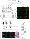
Autophagy activation is mediated by accumulation of ATF4. (A) HeLa cells were either transfected with nontargeting siRNA (Scr.) or with a mix of PSMD4 and ADRM1 siRNAs. In parallel, cells were treated with 2 µg/ml tunicamycin or 2.5 µM velcade overnight. Next, the cells were lysed with NP-40, and lysates were analyzed by western blot using anti-ATF4, anti-DDIT3 and anti-Actin antibodies. (B) Total RNA of HeLa cells transfected with the indicated siRNAs was extracted and subjected to quantitative RT-PCR analysis using SQSTM1 and HPRT1 primers. The level of mRNA was normalized to the HPRT1 level. The results are the mean ± STDEV of at least 3 independent experiments. *p < 0.05 for the difference between treatments and the PSMD4 and ADRM1 siRNA, determined by ANOVA with Dunnet Multiple Comparison Test as post-test. Scr. SiRNA is represented by a dashed line at 1. (C) HeLa cells were transfected with HA-ATF4 for 24 h. The cells were fixed, immunostained with anti-SQSTM1, anti-HA antibodies and DAPI, and analyzed by confocal microscopy. Scale bar: 10 μm. The number of SQSTM1 dots was quantified in the nontransfected and HA-ATF4-transfected cells. The results are the mean ± STDEV of at least 3 independent experiments. *P < 0.05 for the difference between nontransfected and HA-ATF4-transfected cells as determined by paired t-test. (D) To inhibit proteasomal or lysosomal function in D2 and 70z cells, 2.5 µM Velcade or 0.1 µM BafA were added respectively for 8 h. Then, cells were lysed with NP-40 and analyzed by western blot with anti-Ub, anti-SQSTM1, anti-LC3, anti-ATF4 and anti-Actin antibodies. (E) D2 and 70z cells were incubated with or without 0.1 µM BafA for 8 h and cell viability was assessed using propidium iodide staining. The number of dead cells was quantified by FACS. The results are the mean ± STDEV of at least 3 independent experiments. *P < 0.001 for the difference between 70z and D2 dead cells after BafA treatment was determined by paired t-test. (F) Schematic model underlining a possible mechanism for upregulation of SQSTM1-dependent autophagy upon slowdown of proteasomal processing.

Similar articles

Cited by

References

    1. Lamark T, Johansen T. Autophagy: Links with the proteasome. Curr Opin Cell Biol. 2010;22:192-8. https://doi.org/10.1016/j.ceb.2009.11.002. PMID:19962293. - DOI - PubMed
    1. Nedelsky NB, Todd PK, Taylor JP. Autophagy and the ubiquitin-proteasome system: Collaborators in neuroprotection. Biochim Biophys Acta. 2008;1782:691-9. https://doi.org/10.1016/j.bbadis.2008.10.002. PMID:18930136. - DOI - PMC - PubMed
    1. Shaid S, Brandts CH, Serve H, Dikic I. Ubiquitination and selective autophagy. Cell Death Differ. 2013;20:21-30. https://doi.org/10.1038/cdd.2012.72. PMID:22722335. - DOI - PMC - PubMed
    1. Besche HC, Peth A, Goldberg AL. Getting to first base in proteasome assembly. Cell. 2009;138:25-8. https://doi.org/10.1016/j.cell.2009.06.035. PMID:19596233. - DOI - PMC - PubMed
    1. Deveraux Q, Ustrell V, Pickart C, Rechsteiner M. A 26 S protease subunit that binds ubiquitin conjugates. J Biol Chem. 1994;269:7059-61. https://doi.org/10.1128/MCB.16.11.6020. PMID:8125911. - DOI - PubMed