. 2017 Jul 3;6(3):250-258.
doi: 10.1080/21623945.2017.1356505. Epub 2017 Jul 20.

Intracellular lipid droplets support osteoblast function

Affiliations

Intracellular lipid droplets support osteoblast function

Elizabeth Rendina-Ruedy et al. Adipocyte. .

Abstract

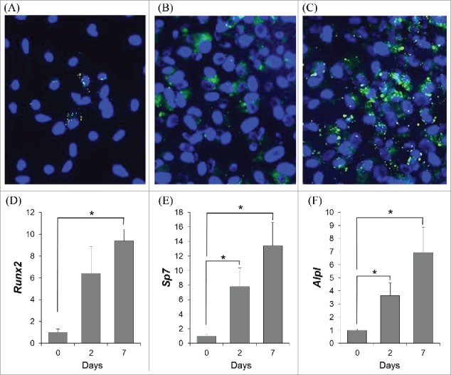
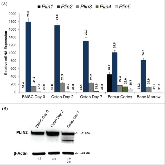
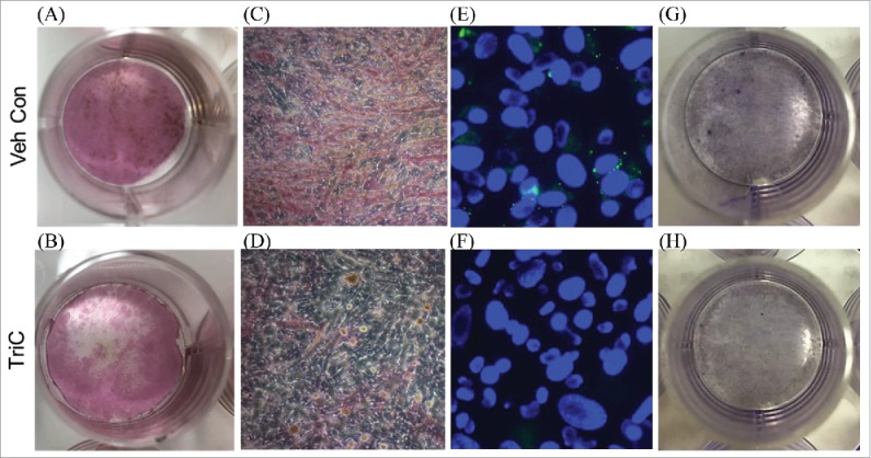
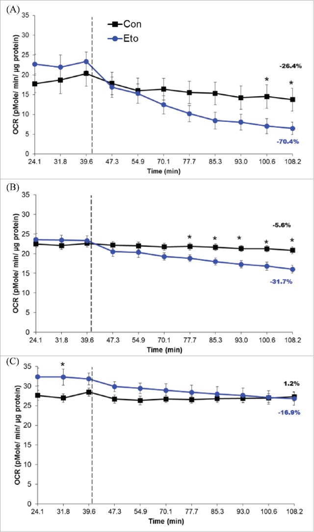
Bone formation is an osteoblast-specific process characterized by high energy demands due to the secretion of matrix proteins and mineralization vesicles. While glucose has been reported as the principle fuel source for osteoblasts, recent evidence supports the tenet that osteoblasts can utilize fatty acids as well. Although the ability to accumulate lipid droplets has been demonstrated in many cell types, there has been little evidence that osteoblasts possess this characteristic. The current study provides evidence that osteoblastogenesis is associated with lipid droplet accumulation capable of supplying energy substrates (fatty acids) required for the differentiation process. Understanding the role of fatty acids in metabolic programming of the osteoblast may lead to novel approaches to increase bone formation and ultimately bone mass.

Keywords: bone; fatty acids; lipolysis; lipophagy; marrow adipocytes.


Figures

Figure 1.
Figure 1.
To determine whether lipid droplets were present during osteoblast differentiation, neutral lipids were selectively stained with BODIPY493/503 (green puncta), while nuclei were stained with Hoechst (blue) in bone marrow stromal cells (BMSCs) under osteogenic conditions for 0 (A), 2 (B), or 7 (C) days; representative images are shown. To confirm osteogenic induction, relative mRNA expression of osteoblast-related genes runt-related transcript factor (Runx2) (D), osterix (Sp7) (E), and alkaline phosphatase (Alpl) (F) were determined in BMSCs through osteogenic differentiation (0, 2, or 7 days). Data shown is presented as mean ± standard error. Symbol, *, denotes statistically significant differences (P < 0.05) between groups.
Figure 2.
Figure 2.
(A) Relative mRNA expression of lipid droplet-associated proteins from the PAT family of proteins including perilipin or Plin1 (Plin1), adipose differentiation-related protein or ADRP (Plin2), tail-differentiation interacting protein of 47 kDa or TIP47 (Plin 3), S3–12 (Plin4), and lipid storage droplet protein 5 or LSDP5 (Plin5) were detected in bone marrow stromal cells (BMSCs) under osteogenic conditions for 0, 2, or 7 days, femur cortex, and the bone marrow. All data are normalized to the housekeeping gene Hprt, and expressed relative to Plin1 expression in day 0 BMSCs. Data is represented as mean ± standard error. Uncorrected, mean CQ values are also indicated on graph for each target gene. (B) Plin2 protein abundance from BMSCs differentiated under osteogenic conditions for 0, 2, and 7 d. Mean protein abundance is expressed as density light units (DLUs x 103) relative to the loading control, β-actin.
Figure 3.
Figure 3.
Primary bone marrow stromal cells (BMSCs) were treated with vehicle (A, C) or TriC (B, D) during the first 24 hours of osteogenic differentiation; treatments were then removed and cells were allowed to differentiate an additional 7 days, and stained for alkaline phosphatase (ALP) and Von Kossa. To confirm TriC's ability to blunt lipid droplet formation, BMSCs were stained with BODIPY493/503 and Hoechst immediately following the initial 24 hour treatment period of vehicle (E) or TriC (F). Additionally, vehicle (G) or TriC (H) treated BMSCs were stained with hematoxylin to confirm cell viability following the initial 24 hours of treatment.
Figure 4.
Figure 4.
Oxygen consumption rates (OCR) from bone marrow stromal cells (BMSCs) cultured under osteogenic conditions for 0 (A), 2 (B), or 7 (C) days. Hashed lines represent time at which XF Base medium (control, Con) or 50 µM etomoxir (Eto) were injected. Percent change in OCR, from baseline to final reading, is also represented on graphs. Data shown is presented as mean ± standard error. Symbol, *, denotes statistically significant differences (P < 0.05) between groups at a given time point.

Similar articles

Cited by

References

    1. Matkovic V, Jelic T, Wardlaw GM, Ilich JZ, Goel PK, Wright JK, Andon MB, Smith KT, Heaney RP. Timing of peak bone mass in Caucasian females and its implication for the prevention of osteoporosis. Inference from a cross-sectional model. J Clin Invest. 1994;93(2):799-08. https://doi.org/10.1172/JCI117034; PMID:8113412 - DOI - PMC - PubMed
    1. Baxter-Jones AD, Faulkner RA, Forwood MR, Mirwald RL, Bailey DA. Bone mineral accrual from 8 to 30 years of age: An estimation of peak bone mass. J Bone Miner Res. 2011;26(8):1729-39. https://doi.org/10.1002/jbmr.412; PMID:21520276 - DOI - PubMed
    1. Borle AB, Nichols N, Nichols G Jr. Metabolic studies of bone in vitro. II. The metabolic patterns of accretion and resorption. J Biol Chem. 1960;235:2351211-1214. PMID:13802862 - PubMed
    1. Borle AB, Nichols N, Nichols G Jr. Metabolic studies of bone in vitro. I. Normal bone. J Biol Chem. 1960;235:2351206-1210. PMID:13802861 - PubMed
    1. Adamek G, Felix R, Guenther HL, Fleisch H. Fatty acid oxidation in bone tissue and bone cells in culture. Characterization and hormonal influences. Biochem J. 11-15-1987;248(1):129-37. https://doi.org/10.1042/bj2480129; PMID:3325035 - DOI - PMC - PubMed

LinkOut - more resources