. 2017 Aug 8;20(6):1360-1371.
doi: 10.1016/j.celrep.2017.07.024.

Peripheral Elevation of a Klotho Fragment Enhances Brain Function and Resilience in Young, Aging, and α-Synuclein Transgenic Mice

Affiliations

Peripheral Elevation of a Klotho Fragment Enhances Brain Function and Resilience in Young, Aging, and α-Synuclein Transgenic Mice

Julio Leon et al. Cell Rep. .

Abstract

Cognitive dysfunction and decreased mobility from aging and neurodegenerative conditions, such as Parkinson and Alzheimer diseases, are major biomedical challenges in need of more effective therapies. Increasing brain resilience may represent a new treatment strategy. Klotho, a longevity factor, enhances cognition when genetically and broadly overexpressed in its full, wild-type form over the mouse lifespan. Whether acute klotho treatment can rapidly enhance cognitive and motor functions or induce resilience is a gap in our knowledge of its therapeutic potential. Here, we show that an α-klotho protein fragment (αKL-F), administered peripherally, surprisingly induced cognitive enhancement and neural resilience despite impermeability to the blood-brain barrier in young, aging, and transgenic α-synuclein mice. αKL-F treatment induced cleavage of the NMDAR subunit GluN2B and also enhanced NMDAR-dependent synaptic plasticity. GluN2B blockade abolished αKL-F-mediated effects. Peripheral αKL-F treatment is sufficient to induce neural enhancement and resilience in mice and may prove therapeutic in humans.

Keywords: Alzheimer disease; NMDA receptors; Parkinson disease; aging; brain; cognition; klotho; mice; motor function; α-synuclein.


Figures

Figure 1
Figure 1. αKL-F Is a Recombinant Post-translationally Modified Fragment of Klotho, and Its Peripheral Administration Does Not Show BBB Crossing or Alteration of Endogenous Hippocampal Klotho Levels
(A) Amino acid sequence of the endogenous α-klotho protein (top, aa 1–1014) and the recombinant α-klotho fragment (αKL-F, bottom, aa 35–982) followed by a His tag. N-T, N terminus; TR, transmembrane region; IC, intracellular domain. (B) Locations of O-glycosylation and N-glycosylation sites of the recombinant protein made in Chinese hamster ovary (CHO) cells. (C) αKL-F was not observed to cross into the brain 4 hr following i.p. injection of Veh or αKL-F (100 μg/kg). Western blot shows immunoprecipitation against His-tagged αKL-F (His-IP) in kidney (detected) and brain (not detected) lysates. αKL-F was added to the veh-treated lysates as a positive control for detection of His-tagged protein (His-Total). (D) Representative western blot showing Klotho and GAPDH levels in homogenized whole hippocampus 4–5 hr following Veh or αKL-F (i.p., 10 μg/kg) treatment (n = 12/group; male; age, 4 months). Images were captured from the same gel. (E) Quantitation of endogenous brain klotho levels showing no differences between Veh- and αKL-F-treated mice. NTG levels are arbitrarily defined as 1.0. Data are mean ± SEM.
Figure 2
Figure 2. αKL-F, Delivered Peripherally, Acutely Enhances Cognition in Young Mice
(A) Diagram of the experimental paradigm of Veh or αKL-F injection (i.p., 10 μg/kg) followed by spatial and working memory testing in the Morris water maze. Mice received daily treatment for 5 days, 16 or 4 hr prior to training and testing (n = 11–18/ group; sex-balanced; age, 4 months). (B) Spatial learning curves (platform hidden) in the water maze. Data represent the daily average of distance traveled to the platform. αKL-F decreased the distance, indicating enhanced learning. Rank-sum test, αKL-F effect days 2–4, p < 0.05. (C) Probe trial with the platform removed 24 hr after completion of hidden training. αKL-F increased the percentage of time spent in the target quadrant, indicating enhanced recall of spatial memory. (D) Probe trial at 24 hr. αKL-F increased the duration of time spent at target, indicating enhanced recall of spatial memory. (E) Velocity in the water maze did not differ between mice treated with Veh or αKL-F. (F) The distance traveled to find a cued, visible platform in the water maze did not differ between mice treated with Veh or αKL-F. (G–I) Diagram of the experimental paradigm of Veh or αKL-F injection (G, i.p., 10 μg/kg), followed by working memory testing in the small Y-maze (H and I). Mice (age, 4 months) received treatment 4 hr prior to testing. (H) Percentages of alternations among arms during exploration of the Y-maze. αKL-F increased alternations, indicating enhanced working memory. In this cohort, mice (males, n = 12–13/group) were exposed to Veh or αKL-F 2 days before testing. (I) Percentages of alternations among arms during exploration of the Y-maze. In a replicate independent cohort, αKL-F increased working memory. Mice (males, n = 9/group) were naive to previous treatments or testing. *p < 0.05, **p < 0.01 (t test). Data are mean ± SEM. See also Figure S1.
Figure 3
Figure 3. αKL-F, Delivered Peripherally, Acutely Enhances Cognition in Aged Mice
(A) Diagram of the experimental paradigm of Veh or αKL-F treatment (i.p., 10 μg/kg) and testing of aged mice in the two-trial Y-maze. (n = 8–10 mice/ group; sex-balanced groups; age, 18 months). Mice received an injection followed by training (24 hr after treatment) and then testing (40 hr after treatment). (B) Duration of time spent in the novel compared with the familiar arm, expressed as a ratio to indicate novel arm preference. αKL-F increased spatial and working memory in aged mice over time. Two-way repeated measures ANOVA, αKL-F effect, **p < 0.01. Data are mean ± SEM.
Figure 4
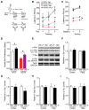
Figure 4. αKL-F, Delivered Peripherally, Induces Neural Resilience by Acutely Countering Cognitive and Motor Deficits in Trans-genic hSYN Mice
(A) Diagram of the experimental paradigm of Veh or αKL-F treatment (i.p., 2.5 μg/kg). Mice received single daily injections for 6 days. On day 4, NTG and hSYN mice were tested on the rotarod to assess motor performance. On day 7 (17 hr after the last injection), mice were tested in the two-trial Y-maze to assess spatial and working cognition. (B) Motor learning and function measured by time on the rotarod during training. hSYN mice showed motor dysfunction compared with NTG mice. αKL-F treatment improved motor learning in hSYN mice (n = 10–15 mice/group; male; age, 3–6 months). Mixed model ANVOA: hSYN effect, p < 0.0001. (C) Motor function measured by time on the rotarod during testing. hSYN mice showed motor dysfunction compared with NTG mice. Mixed model ANOVA: hSYN effect, p < 0.0001. αKL-F treatment increased time on the rotarod across sessions in hSYN mice. Two-way repeated measures ANOVA: αKL-F effect, *p < 0.05 as indicated by brackets. (D) Duration of time spent in the novel compared with the familiar arm during testing, expressed as a ratio to indicate novel arm preference at 5 min of exploration. αKL-F improved cognitive deficits in hSYN mice (n = 14–23 mice/group; male; age, 3–6 months). Two-way ANOVA: hSYN effect, p < 0.0001; αKL-F effect, p < 0.01. (E–I) The hippocampus and cortex of Veh- and αKL-F-treated hSYN mice were analyzed after 3 days of daily injection (n = 5–6 mice/group; male; age, 3–7 months). (E) Representative western blots showing hippocampal levels of total α-Syn, phosphorylated α-Syn (p-α-Syn, Ser-129), total mouse tau (T-Tau), phosphorylated mouse tau (p-Tau, Ser-396/404), Actin, and GAPDH in Veh- and αKL-F-treated hSYN mice. (F) Quantitation of α-Syn levels, showing no differences between Veh- and αKL-F-treated hSYN mice. (G) Quantitation of p-α-Syn (Ser-129) levels, showing no differences between Veh- and αKL-F-treated hSYN mice. (H) Quantitation of T-Tau levels, showing no differences between Veh- and αKL-F-treated hSYN mice. (I) Quantitation of p-Tau levels, showing no differences between Veh- and αKL-F-treated hSYN mice. #p = 0.10, *p < 0.05 versus trial 1 (B, paired t tests) or as indicated by brackets (C, mean effect; D, unpaired t tests) via Bonferroni-Holm test. Data are mean ± SEM.
Figure 5
Figure 5. αKL-F Does Not Alter Synaptic Levels of HMW GluN2B
(A–C) Synaptic membrane fraction 1 (PSD-enriched) and fraction 2 (non-PSD-enriched) isolated from the hippocampus of mice (age, 4 months; male) 4 hr after a single treatment followed by the small Y-maze or after 5 days of daily treatment with Veh or αKL-F (i.p., 10 μg/kg). (A) Representative western blots from hippocampal membrane fractions from mice 4 hr (top) following a single treatment or 5 days following daily treatments (bottom). Only the HMW GluN2B band was observed. (B) Quantitation of PSD-enriched (fraction 1) GluN2B protein levels 4 hr following treatment (n = 4/group). (C) Quantitation of PSD-enriched (fraction 1) GluN2B protein levels 5 days following daily treatment (n = 10/group). Data are mean ± SEM.
Figure 6
Figure 6. αKL-F Acutely Increases an LMW Form of GluN2B
(A–E) Whole hippocampal lysate isolated from mice treated with Veh or αKL-F (10 μg/kg i.p.) after 4 hr of a single treatment followed by the small Y-maze or 5 days of daily treatment and then assessed for GluN2B levels. (A) Representative western blots from hippocampal total lysates (with 1% SDS) from mice with Veh or αKL-F 4 hr after treatment (top, images were captured from the same gel) or 5 days following daily treatment (bottom) (age, 4 months; male). HMW and LMW GluN2B forms were observed. (B) Quantitation of LMW/total (HMW+LMW) GluN2B protein levels from the hippocampus 4 hr after treatment (n = 10/group). (C) Correlation of relative LMW/total GluN2B fragment levels from mice shown in (D) with percentage alternations in the small Y-maze (R2 = 0.5, p < 0.03 with αKL-F treatment). (D) Replicate independent cohort quantitation of LMW/total GluN2B protein levels from the hippocampus 4 hr after treatment (n = 21/group). (E) Quantitation of LMW/total GluN2B protein levels from the hippocampus following 5 days of daily treatment in the absence of behavior studies (n = 11–12/group). (F) Western blot showing the specificity of the HMW and LMW GluN2B band by immunoprecipitation with and without preincubation with a GluN2B-blocking peptide harboring the GluN2B antibody epitope sequence. #p < 0.10, *p < 0.05 versus Veh as indicated by brackets. Data are mean ± SEM. See also Figure S2.
Figure 7
Figure 7. αKL-F Acutely Enhances NMDAR-Dependent Synaptic Plasticity in the Hippocampus, and Selective Blockade of the NMDAR Subunit GluN2B Abolishes αKL-F Effects on Cognition
(A and B) fEPSP recordings from acute hippocampal slices of 3-month-old male mice (n = 7–14 slices/group, 3 mice/group) treated with Veh or αKL-F 4h prior to brain slicing. (A) LTP induction and potentiation in the CA1 region was monitored for 30 min following theta burst stimulation of the Schaffer collateral pathway. (B) Average fEPSP slope averaged over 30 min in hippocampal slices of mice treated with Veh or αKL-F. Two-way repeated measures ANOVA: αKL-F effect, p < 0.01. (C and D) Mice (n = 15/group; male; age, 4 months) were treated i.p. with Veh or αKL-F (10 μg/kg) 4 hr prior to testing, followed by i.p. saline (−) or Ro 25 (+) (5 mg/kg) 10 min prior to testing in the small Y-maze. (C) Percentages of alternations among arms during exploration of the small Y-maze over 4 min. αKL-F increased alternations, and Ro 25 preferentially decreased αKL-F-mediated effects. Two-way ANOVA: Ro 25 by αKL-F interaction, p < 0.05. (D) Percentage of decrease in alternations following Ro 25 treatment in Veh-and αKL-F-treated mice. Ro 25 preferentially decreased the percentage of alternations in mice with αKL-F treatment. *p < 0.05, **p < 0.01 versus Veh or as indicated by brackets by t test (B and D) or Bonferroni-Holm test (C). Data are mean ± SEM. See also Figure S3.

References

    1. Almeida OP, Morar B, Hankey GJ, Yeap BB, Golledge J, Jablensky A, Flicker L. Longevity Klotho gene polymorphism and the risk of dementia in older men. Maturitas. 2017;101:1–5. - PubMed
    1. Alvarez-Castelao B, Schuman EM. The Regulation of Synaptic Protein Turnover. J Biol Chem. 2015;290:28623–28630. - PMC - PubMed
    1. Arking DE, Krebsova A, Macek M, Sr, Macek M, Jr, Arking A, Mian IS, Fried L, Hamosh A, Dey S, McIntosh I, Dietz HC. Association of human aging with a functional variant of klotho. Proc Natl Acad Sci USA. 2002;99:856–861. - PMC - PubMed
    1. Bennett DA. Mixed pathologies and neural reserve: Implications of complexity for Alzheimer disease drug discovery. PLoS Med. 2017;14:e1002256. - PMC - PubMed
    1. Bloch L, Sineshchekova O, Reichenbach D, Reiss K, Saftig P, Kuro-o M, Kaether C. Klotho is a substrate for alpha-, beta- and gamma-secretase. FEBS Lett. 2009;583:3221–3224. - PMC - PubMed

MeSH terms