Nanosized Hydroxyapatite Coating on PEEK Implants Enhances Early Bone Formation: A Histological and Three-Dimensional Investigation in Rabbit Bone
- PMID: 28793409
- PMCID: PMC5455651
- DOI: 10.3390/ma8073815
Nanosized Hydroxyapatite Coating on PEEK Implants Enhances Early Bone Formation: A Histological and Three-Dimensional Investigation in Rabbit Bone
Abstract
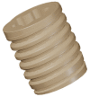
Polyether ether ketone (PEEK) has been frequently used in spinal surgery with good clinical results. The material has a low elastic modulus and is radiolucent. However, in oral implantology PEEK has displayed inferior ability to osseointegrate compared to titanium materials. One idea to reinforce PEEK would be to coat it with hydroxyapatite (HA), a ceramic material of good biocompatibility. In the present study we analyzed HA-coated PEEK tibial implants via histology and radiography when following up at 3 and 12 weeks. Of the 48 implants, 24 were HA-coated PEEK screws (test) and another 24 implants served as uncoated PEEK controls. HA-coated PEEK implants were always osseointegrated. The total bone area (BA) was higher for test compared to control implants at 3 (p < 0.05) and 12 weeks (p < 0.05). Mean bone implant contact (BIC) percentage was significantly higher (p = 0.024) for the test compared to control implants at 3 weeks and higher without statistical significance at 12 weeks. The effect of HA-coating was concluded to be significant with respect to early bone formation, and HA-coated PEEK implants may represent a good material to serve as bone anchored clinical devices.
Keywords: HA; biomaterial; histology; polyether ether ketone.
Conflict of interest statement
The authors declare no conflict of interest.
Figures


References
-
- Branemark P.I., Hansson B.O., Adell R., Breine U., Lindstrom J., Hallen O., Ohman A. Osseointegrated implants in the treatment of the edentulous jaw. Experience from a 10-year period. Scand. J. Plast. Reconstr. Surg. Suppl. 1977;16:1–132. - PubMed
-
- Head W.C., Bauk D.J., Emerson R.H., Jr. Titanium as the material of choice for cementless femoral components in total hip arthroplasty. Clin. Orthop. Relat. Res. 1995:85–90. - PubMed
-
- Treves C., Martinesi M., Stio M., Gutierrez A., Jimenez J.A., Lopez M.F. In vitro biocompatibility evaluation of surface-modified titanium alloys. J. Biomed. Mater. Res. Part A. 2010;92:1623–1634. - PubMed
LinkOut - more resources
Full Text Sources
Other Literature Sources

