. 2017 Oct 1;130(19):3222-3233.
doi: 10.1242/jcs.201715. Epub 2017 Aug 9.

Iron affects Ire1 clustering propensity and the amplitude of endoplasmic reticulum stress signaling

Affiliations

Iron affects Ire1 clustering propensity and the amplitude of endoplasmic reticulum stress signaling

Nir Cohen et al. J Cell Sci. .

Abstract

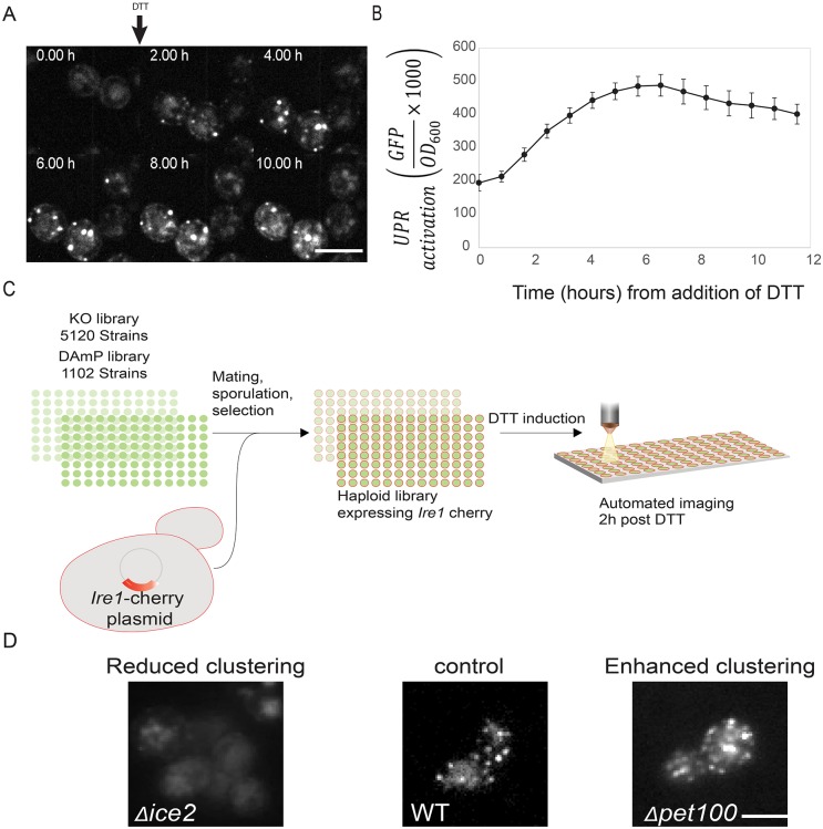
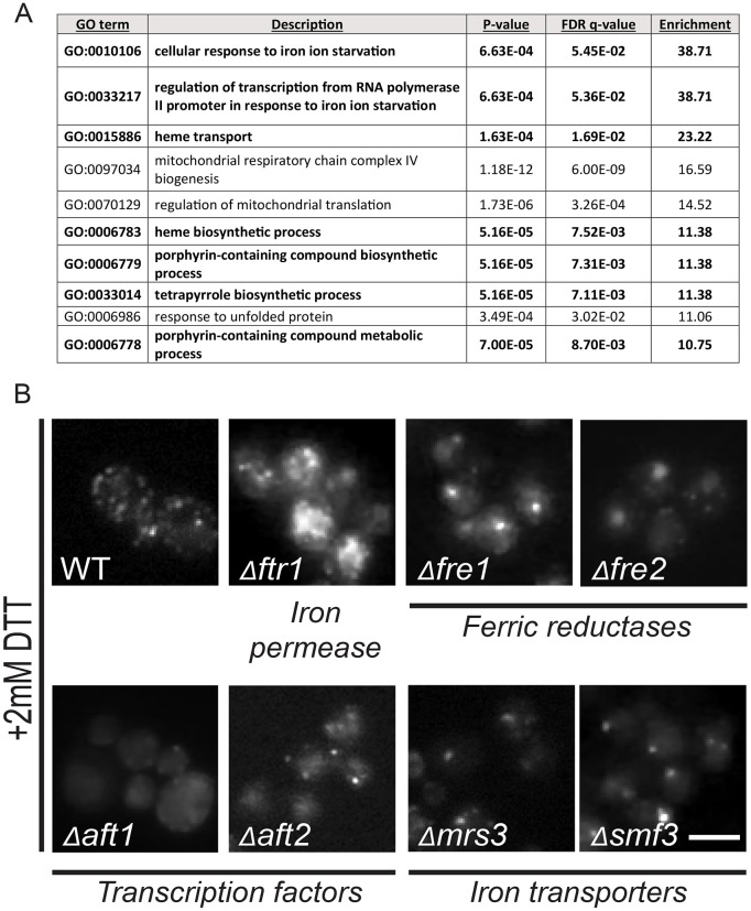
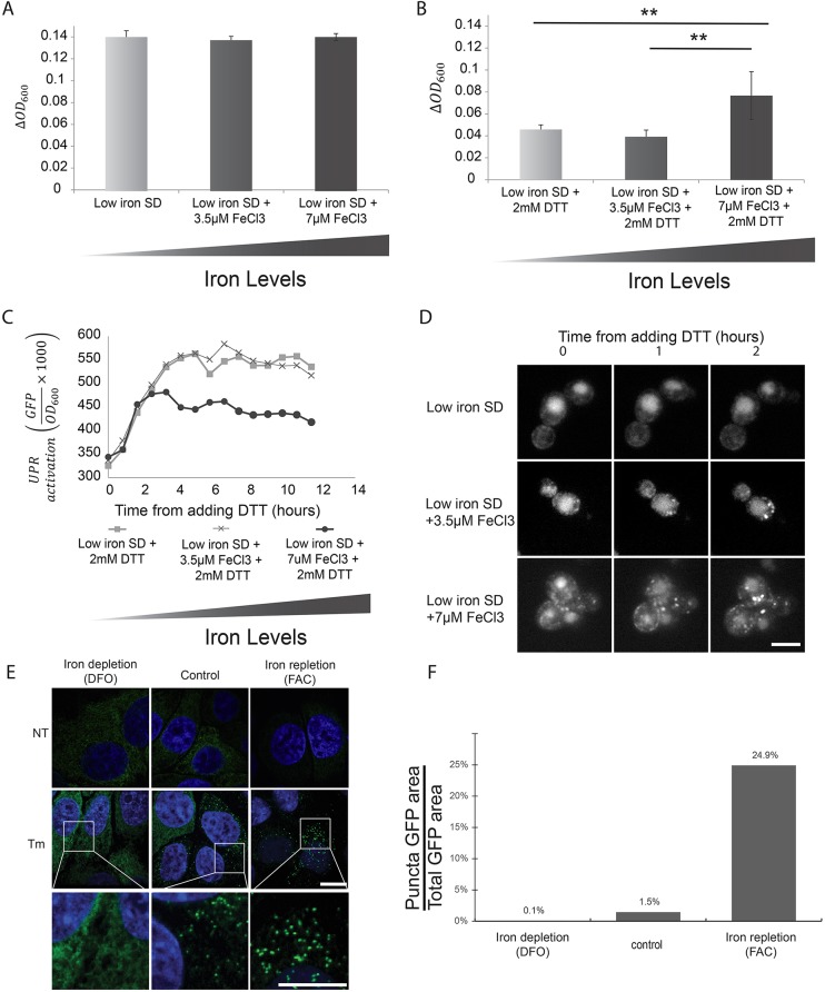
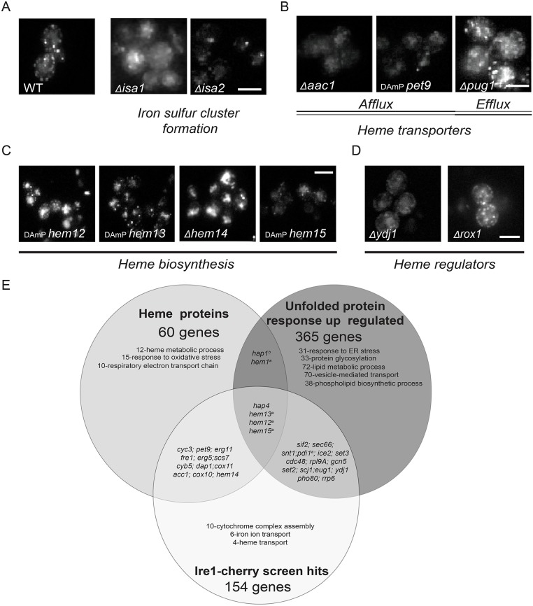
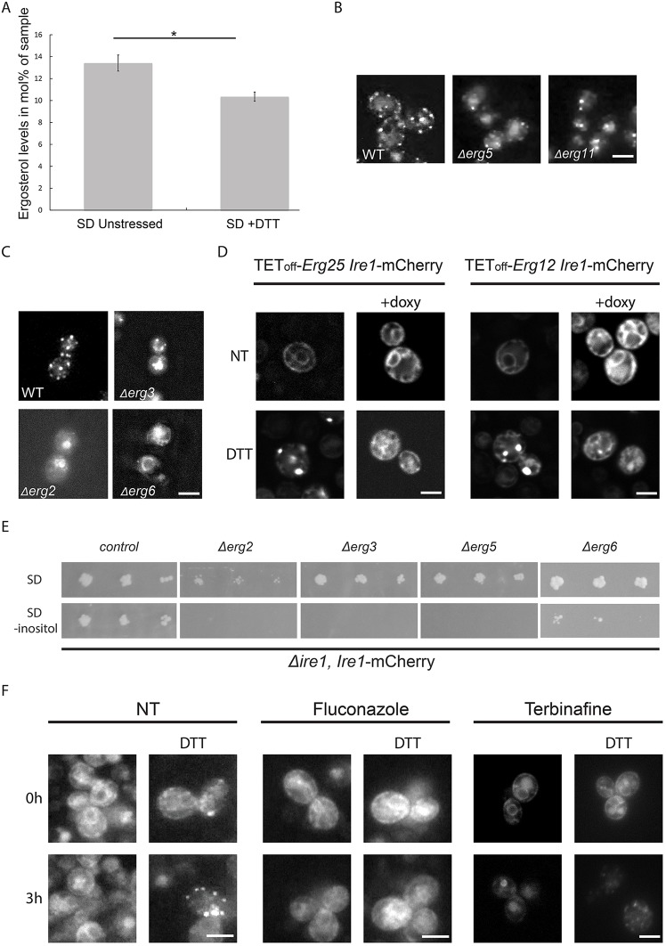
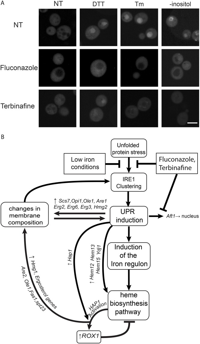
The unfolded protein response (UPR) allows cells to adjust secretory pathway capacity according to need. Ire1, the endoplasmic reticulum (ER) stress sensor and central activator of the UPR is conserved from the budding yeast Saccharomyces cerevisiae to humans. Under ER stress conditions, Ire1 clusters into foci that enable optimal UPR activation. To discover factors that affect Ire1 clustering, we performed a high-content screen using a whole-genome yeast mutant library expressing Ire1-mCherry. We imaged the strains following UPR induction and found 154 strains that displayed alterations in Ire1 clustering. The hits were enriched for iron and heme effectors and binding proteins. By performing pharmacological depletion and repletion, we confirmed that iron (Fe3+) affects UPR activation in both yeast and human cells. We suggest that Ire1 clustering propensity depends on membrane composition, which is governed by heme-dependent biosynthesis of sterols. Our findings highlight the diverse cellular functions that feed into the UPR and emphasize the cross-talk between organelles required to concertedly maintain homeostasis.

Keywords: Heme; Ire1; Iron; Saccharomyces cerevisiae; Sterol; UPR.


Conflict of interest statement

Competing interestsThe authors declare no competing or financial interests.

Figures

Fig. 1.
Fig. 1.
A systematic high-content screen reveals effectors of Ire1 clustering. (A) Ire1–mCherry was visualized over time following induction of the UPR by 2 mM DTT and shows clustering as expected. (B) UPR activity was measured over time using a UPRE-GFP reporter following induction of UPR by 2 mM DTT. UPR activity increases until an activation plateau is reached. Results are mean±s.d. (n=9). (C) Work flow for systematic screen to uncover Ire1–mCherry clustering effectors. (D) Hits from the Ire1–mCherry screen were divided into two phenotypic groups. WT, wild type. Scale bars: 5 μm.
Fig. 2.
Fig. 2.
Iron homeostasis affects Ire1–mCherry clustering. (A) GO term enrichment of the hits in the screen based on the GOrilla analysis website. The table shows the ten highest enriched categories excluding redundant GO terms. In bold are all iron- and heme-related processes. (B) Deletion of several iron homeostasis genes disrupts Ire1–mCherry clustering following ER stress (2 mM DTT) (images from original screen). WT, wild type. Scale bar: 5 μm.
Fig. 3.
Fig. 3.
Iron is essential for mounting a productive UPR. (A) Iron depletion or repletion does not affect yeast growth under non-ER stress conditions as assayed by the change of OD600 per hour using a plate reader. (B) Iron repletion is essential to enable robust yeast growth during ER stress (2 mM DTT) as assayed by the change of OD600 per hour using a plate reader. Results are mean±s.d. (n=9). **P<0.01 (Student's t-test). (C) The presence of iron enables mounting a more effective UPR response as can be seen by measuring the UPRE-GFP reporter over time under various iron repletion or depletion conditions following ER stress (2 mM DTT). (D) Ire1–mCherry clustering following induction of ER stress (2 mM DTT) is affected by the amount of iron in the medium. Scale bar: 5 μm. (E) Iron affects Ire1 clustering in mammalian HeLa cells treated with 10 µg/ml tunicamycin (Tm). While iron depletion (100 µM DFO) disrupts Ire1–GFP clustering, iron repletion (300 µM FAC) enhances it. The bottom row shows a magnification of the Tm-treated cells. Scale bar: 10 μm. (F) Quantification of results in E for DFO=146, FAC=156 and control=135 cells. NT, not treated; WT, wild type.
Fig. 4.
Fig. 4.
ISC and heme biosynthesis are essential for Ire1–mCherry clustering. (A) Deletion of the ISC biosynthesis proteins, Δisa1 and Δisa2 abrogates Ire1–mCherry clustering following stress (images from original screen). (B) Reducing heme afflux (Δacc1, pet9-DAmP) disrupts Ire1–mCherry clustering following stress, while reducing heme efflux (Δpug1) enhances it (images from original screen). (C) Mutations of the enzymes required for the last steps of the heme biosynthesis pathway disrupt Ire1–mCherry clustering following stress (images from original screen). (D) Deletion of Ydj1, the positive regulator of the heme-specific transcription factor HAP1, disrupts Ire1-mCherry clustering following stress, while deletion of the HAP1 negative regulator, Rox1, enhances it (images from original screen). (E) Venn diagram overlaying heme-containing proteins, UPR upregulated genes and Ire1 clustering effectors from the screen (only the major GO term enrichments are shown for each group). The diagram highlights a potential role for ergosterol biosynthesis (a, hypomorphic allele; b, not in the screen). WT, wild type. Scale bars: 5 μm.
Fig. 5.
Fig. 5.
Ergosterol biosynthesis affects Ire1-mCherry clustering. (A) ER stress causes a rapid and significant decrease in overall ergosterol levels. Analysis of the ergosterol content is shown in unstressed cells and following 2 mM DTT treatment for 1 h. Results are mean±s.d. (n=3). *P<0.05 (Student's t-test). (B) Deletions of the heme-containing ergosterol biosynthesis pathway enzymes Erg5 and Erg11 disrupt Ire1–mCherry clustering following ER stress (images from original screen). (C) Deletions of the non-essential ergosterol biosynthesis pathway enzymes, Erg2, Erg3 and Erg6, disrupt Ire1–mCherry clustering following ER stress (2 mM DTT). (D) Repressing the expression of the two essential ergosterol biosynthesis pathway enzymes Erg12 and Erg25 using a TetOFF promotor repressed by doxycycline disrupts Ire1–mCherry clustering following ER stress (2 mM DTT). (E) Deletion strains of the non-essential components of the ergosterol biosynthesis pathway cannot grow even in mild ER stress conditions caused by removal of inositol from the medium (SD –inositol). (F) Short-term inhibition of ergosterol biosynthesis using two different inhibitors (20 μg/ml Fluconazole or 10 μg/ml Terbinafine) disrupts Ire1–mCherry clustering following UPR induction (2 mM DTT). NT, not treated; WT, wild type. Scale bars: 5 μm.
Fig. 6.
Fig. 6.
Suggested cross-talk between the UPR, heme and iron homeostatic machinery. (A) GFP–Aft1 re-localizes to the nucleus during unfolded protein stress, as induced by either 2 mM DTT, 5 μg/ml tunicamycin or 24 h growth in inositol depletion medium. The nuclear accumulation is inhibited by lowering ergosterol biosynthesis by using ergosterol biosynthesis inhibitors (Fluconazole 20 μg/ml or Terbinafine 10 μg/ml). NT, not treated. Scale bar: 5 μm. (B) A schematic for a hypothetical feedback loop suggested for the heme and iron cross-talk with ergosterol biosynthesis and the UPR.

Similar articles

Cited by

References

    1. Abraham N. G., Tsenovoy P. L., McClung J. and Drummond G. S. (2008). Heme oxygenase: a target gene for anti-diabetic and obesity. Curr. Pharm. Des. 14, 412-421. 10.2174/138161208783597371 - DOI - PubMed
    1. Aragón T., van Anken E., Pincus D., Serafimova I. M., Korennykh A. V., Rubio C. A. and Walter P. (2009). Messenger RNA targeting to endoplasmic reticulum stress signalling sites. Nature 457, 736-740. 10.1038/nature07641 - DOI - PMC - PubMed
    1. Breker M., Gymrek M. and Schuldiner M. (2013). A novel single-cell screening platform reveals proteome plasticity during yeast stress responses. J. Cell Biol. 200, 839-850. 10.1083/jcb.201301120 - DOI - PMC - PubMed
    1. Brachmann C.B., Davies A., Cost G.J., Caputo E., Li J., Hieter P. and Boeke J.D. (1998). Designer deletion strains derived from Saccharomyces cerevisiae S288C: a useful set of strains and plasmids for PCR-mediated gene disruption and other applications. Yeast 14, 115-132. 10.1002/(SICI)1097-0061(19980130)14:2<115::AID-YEA204>3.0.CO;2-2 - DOI - PubMed
    1. Breslow D. K., Cameron D. M., Collins S. R., Schuldiner M., Stewart-Ornstein J., Newman H. W., Braun S., Madhani H. D., Krogan N. J. and Weissman J. S. (2008). A comprehensive strategy enabling high-resolution functional analysis of the yeast genome. Nat. Methods 5, 711-718. 10.1038/nmeth.1234 - DOI - PMC - PubMed

MeSH terms